4XOU

Crystal structure of the SR Ca2+-ATPase in the Ca2-E1-MgAMPPCP form determined by serial femtosecond crystallography using an X-ray free-electron laser.


Experimental Data Snapshot

  • Method: X-RAY DIFFRACTION
  • Resolution: 2.80 Å
  • R-Value Free: 0.343 
  • R-Value Work: 0.304 
  • R-Value Observed: 0.305 

wwPDB Validation   3D Report Full Report


Ligand Structure Quality Assessment 


This is version 1.6 of the entry. See complete history


Literature

Structural studies of P-type ATPase-ligand complexes using an X-ray free-electron laser.

Bublitz, M.Nass, K.Drachmann, N.D.Markvardsen, A.J.Gutmann, M.J.Barends, T.R.Mattle, D.Shoeman, R.L.Doak, R.B.Boutet, S.Messerschmidt, M.Seibert, M.M.Williams, G.J.Foucar, L.Reinhard, L.Sitsel, O.Gregersen, J.L.Clausen, J.D.Boesen, T.Gotfryd, K.Wang, K.T.Olesen, C.Moller, J.V.Nissen, P.Schlichting, I.

(2015) IUCrJ 2: 409-420

  • DOI: https://doi.org/10.1107/S2052252515008969
  • Primary Citation of Related Structures:  
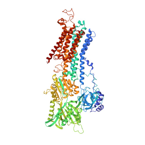
    4XOU

  • PubMed Abstract: 

    Membrane proteins are key players in biological systems, mediating signalling events and the specific transport of e.g. ions and metabolites. Consequently, membrane proteins are targeted by a large number of currently approved drugs. Understanding their functions and molecular mechanisms is greatly dependent on structural information, not least on complexes with functionally or medically important ligands. Structure determination, however, is hampered by the difficulty of obtaining well diffracting, macroscopic crystals. Here, the feasibility of X-ray free-electron-laser-based serial femtosecond crystallography (SFX) for the structure determination of membrane protein-ligand complexes using microcrystals of various native-source and recombinant P-type ATPase complexes is demonstrated. The data reveal the binding sites of a variety of ligands, including lipids and inhibitors such as the hallmark P-type ATPase inhibitor orthovanadate. By analyzing the resolution dependence of ligand densities and overall model qualities, SFX data quality metrics as well as suitable refinement procedures are discussed. Even at relatively low resolution and multiplicity, the identification of ligands can be demonstrated. This makes SFX a useful tool for ligand screening and thus for unravelling the molecular mechanisms of biologically active proteins.


  • Organizational Affiliation

    Department of Molecular Biology and Genetics, Centre for Membrane Pumps in Cells and Disease - PUMPkin, Danish National Research Foundation, Aarhus University , Gustav Wieds Vej 10c, 8000 Aarhus C, Denmark.


Macromolecules
Find similar proteins by:  (by identity cutoff)  |  3D Structure
Entity ID: 1
MoleculeChains Sequence LengthOrganismDetailsImage
Sarcoplasmic/endoplasmic reticulum calcium ATPase 1994Oryctolagus cuniculusMutation(s): 0 
EC: 3.6.3.8
Membrane Entity: Yes 
UniProt
Find proteins for P04191 (Oryctolagus cuniculus)
Explore P04191 
Go to UniProtKB:  P04191
Entity Groups  
Sequence Clusters30% Identity50% Identity70% Identity90% Identity95% Identity100% Identity
UniProt GroupP04191
Sequence Annotations
Expand
  • Reference Sequence
Experimental Data & Validation

Experimental Data

  • Method: X-RAY DIFFRACTION
  • Resolution: 2.80 Å
  • R-Value Free: 0.343 
  • R-Value Work: 0.304 
  • R-Value Observed: 0.305 
  • Space Group: C 1 2 1
Unit Cell:
Length ( Å )Angle ( ˚ )
a = 162α = 90
b = 76.3β = 109
c = 151.1γ = 90
Software Package:
Software NamePurpose
PHENIXrefinement
PHASERphasing
PDB_EXTRACTdata extraction
CASSdata processing
CrystFELdata processing

Structure Validation

View Full Validation Report


Ligand Structure Quality Assessment 


Entry History & Funding Information

Deposition Data


Funding OrganizationLocationGrant Number
Department of Energy (DOE, United States)United States--
Danish Council for Independent ResearchDenmark0602-02495b
Danish National Research FoundationDenmark--
European Research CouncilDenmark--

Revision History  (Full details and data files)

  • Version 1.0: 2015-06-10
    Type: Initial release
  • Version 1.1: 2015-07-29
    Changes: Database references
  • Version 1.2: 2017-08-09
    Changes: Author supporting evidence, Database references
  • Version 1.3: 2017-09-13
    Changes: Author supporting evidence
  • Version 1.4: 2018-11-14
    Changes: Data collection
  • Version 1.5: 2022-03-30
    Changes: Author supporting evidence, Database references, Derived calculations
  • Version 1.6: 2024-01-10
    Changes: Data collection, Refinement description