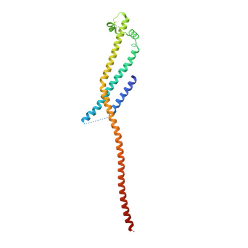
Integrin Engagement by the Helical RGD Motif of the Helicobacter pylori CagL Protein Is Regulated by pH-induced Displacement of a Neighboring Helix.
Bonsor, D.A., Pham, K.T., Beadenkopf, R., Diederichs, K., Haas, R., Beckett, D., Fischer, W., Sundberg, E.J.(2015) J Biol Chem 290: 12929-12940
- PubMed: 25837254
- DOI: https://doi.org/10.1074/jbc.M115.641829
- Primary Citation of Related Structures:
4X5U - PubMed Abstract:
Arginine-aspartate-glycine (RGD) motifs are recognized by integrins to bridge cells to one another and the extracellular matrix. RGD motifs typically reside in exposed loop conformations. X-ray crystal structures of the Helicobacter pylori protein CagL revealed that RGD motifs can also exist in helical regions of proteins. Interactions between CagL and host gastric epithelial cell via integrins are required for the translocation of the bacterial oncoprotein CagA. Here, we have investigated the molecular basis of the CagL-host cell interactions using structural, biophysical, and functional analyses. We solved an x-ray crystal structure of CagL that revealed conformational changes induced by low pH not present in previous structures. Using analytical ultracentrifugation, we found that pH-induced conformational changes in CagL occur in solution and not just in the crystalline environment. By designing numerous CagL mutants based on all available crystal structures, we probed the functional roles of CagL conformational changes on cell surface integrin engagement. Together, our data indicate that the helical RGD motif in CagL is buried by a neighboring helix at low pH to inhibit CagL binding to integrin, whereas at neutral pH the neighboring helix is displaced to allow integrin access to the CagL RGD motif. This novel molecular mechanism of regulating integrin-RGD motif interactions by changes in the chemical environment provides new insight to H. pylori-mediated oncogenesis.
Organizational Affiliation:
From the Institute of Human Virology and.