4WW7

Crystal structure of binary complex Bud32-Cgi121 in complex with AMP


Experimental Data Snapshot

  • Method: X-RAY DIFFRACTION
  • Resolution: 1.67 Å
  • R-Value Free: 0.213 
  • R-Value Work: 0.193 
  • R-Value Observed: 0.194 

wwPDB Validation   3D Report Full Report


Ligand Structure Quality Assessment 


This is version 1.1 of the entry. See complete history


Literature

Crystal structures of the Gon7/Pcc1 and Bud32/Cgi121 complexes provide a model for the complete yeast KEOPS complex.

Zhang, W.Collinet, B.Graille, M.Daugeron, M.C.Lazar, N.Libri, D.Durand, D.van Tilbeurgh, H.

(2015) Nucleic Acids Res 43: 3358-3372

  • DOI: https://doi.org/10.1093/nar/gkv155
  • Primary Citation of Related Structures:  
    4WW5, 4WW7, 4WW9, 4WWA, 4WX8, 4WXA, 4XAH

  • PubMed Abstract: 

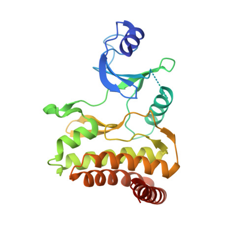
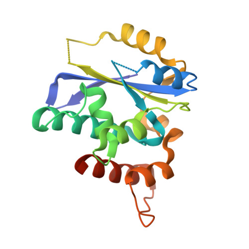
    The yeast KEOPS protein complex comprising Kae1, Bud32, Cgi121, Pcc1 and Gon7 is responsible for the essential tRNA threonylcarbamoyladenosine (t(6)A) modification. Deletion of genes coding for the KEOPS subunits also affects telomere elongation and transcriptional regulation. In the present work, the crystal structure of Bud32/Cgi121 in complex with ADP revealed that ADP is bound in the catalytic site of Bud32 in a canonical manner characteristic of Protein Kinase A (PKA) family proteins. We found that Gon7 forms a stable heterodimer with Pcc1 and report the crystal structure of the Pcc1-Gon7 heterodimer. Gon7 interacts with the same Pcc1 region engaged in the archaeal Pcc1 homodimer. We further show that yeast KEOPS, unlike its archaeal counterpart, exists as a heteropentamer in which Gon7, Pcc1, Kae1, Bud32 and Cgi121 also adopt a linear arrangement. We constructed a model of yeast KEOPS that provides structural insight into the role of Gon7. The model also revealed the presence of a highly positively charged crater surrounding the entrance of Kae1 that likely binds tRNA.


  • Organizational Affiliation

    Institut de Biologie Intégrative de la Cellule, UMR 9198, CNRS, Université de Paris Sud XI, Bâtiment 430, 91405 Orsay, France.


Macromolecules
Find similar proteins by:  (by identity cutoff)  |  3D Structure
Entity ID: 1
MoleculeChains Sequence LengthOrganismDetailsImage
EKC/KEOPS complex subunit BUD32261Saccharomyces cerevisiae S288CMutation(s): 0 
Gene Names: BUD32LDB14YGR262C
EC: 3.6 (PDB Primary Data), 2.7.11.1 (PDB Primary Data)
UniProt
Find proteins for P53323 (Saccharomyces cerevisiae (strain ATCC 204508 / S288c))
Explore P53323 
Go to UniProtKB:  P53323
Entity Groups  
Sequence Clusters30% Identity50% Identity70% Identity90% Identity95% Identity100% Identity
UniProt GroupP53323
Sequence Annotations
Expand
  • Reference Sequence
Find similar proteins by:  (by identity cutoff)  |  3D Structure
Entity ID: 2
MoleculeChains Sequence LengthOrganismDetailsImage
EKC/KEOPS complex subunit CGI121187Saccharomyces cerevisiae S288CMutation(s): 0 
Gene Names: CGI121YML036W
UniProt
Find proteins for Q03705 (Saccharomyces cerevisiae (strain ATCC 204508 / S288c))
Explore Q03705 
Go to UniProtKB:  Q03705
Entity Groups  
Sequence Clusters30% Identity50% Identity70% Identity90% Identity95% Identity100% Identity
UniProt GroupQ03705
Sequence Annotations
Expand
  • Reference Sequence
Small Molecules
Ligands 3 Unique
IDChains Name / Formula / InChI Key2D Diagram3D Interactions
AMP
Query on AMP

Download Ideal Coordinates CCD File 
C [auth A]ADENOSINE MONOPHOSPHATE
C10 H14 N5 O7 P
UDMBCSSLTHHNCD-KQYNXXCUSA-N
SO4
Query on SO4

Download Ideal Coordinates CCD File 
H [auth A],
I [auth A],
J [auth B]
SULFATE ION
O4 S
QAOWNCQODCNURD-UHFFFAOYSA-L
ACT
Query on ACT

Download Ideal Coordinates CCD File 
D [auth A],
E [auth A],
F [auth A],
G [auth A]
ACETATE ION
C2 H3 O2
QTBSBXVTEAMEQO-UHFFFAOYSA-M
Experimental Data & Validation

Experimental Data

  • Method: X-RAY DIFFRACTION
  • Resolution: 1.67 Å
  • R-Value Free: 0.213 
  • R-Value Work: 0.193 
  • R-Value Observed: 0.194 
  • Space Group: I 41
Unit Cell:
Length ( Å )Angle ( ˚ )
a = 113.89α = 90
b = 113.89β = 90
c = 87.41γ = 90
Software Package:
Software NamePurpose
PHENIXrefinement

Structure Validation

View Full Validation Report


Ligand Structure Quality Assessment 


Entry History 

Deposition Data

Revision History  (Full details and data files)

  • Version 1.0: 2015-03-18
    Type: Initial release
  • Version 1.1: 2015-04-15
    Changes: Database references