4WTV

Crystal structure of the phosphatidylinositol 4-kinase IIbeta


Experimental Data Snapshot

  • Method: X-RAY DIFFRACTION
  • Resolution: 1.90 Å
  • R-Value Free: 0.240 
  • R-Value Work: 0.209 
  • R-Value Observed: 0.210 

wwPDB Validation   3D Report Full Report


Ligand Structure Quality Assessment 


This is version 1.2 of the entry. See complete history


Literature

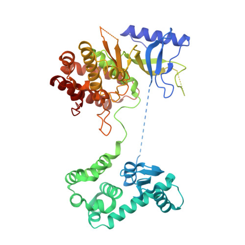
The high-resolution crystal structure of phosphatidylinositol 4-kinase II beta and the crystal structure of phosphatidylinositol 4-kinase II alpha containing a nucleoside analogue provide a structural basis for isoform-specific inhibitor design.

Klima, M.Baumlova, A.Chalupska, D.Hrebabecky, H.Dejmek, M.Nencka, R.Boura, E.

(2015) Acta Crystallogr D Biol Crystallogr 71: 1555-1563

  • DOI: https://doi.org/10.1107/S1399004715009505
  • Primary Citation of Related Structures:  
    4WTV, 4YC4

  • PubMed Abstract: 

    Phosphatidylinositol 4-phosphate (PI4P) is the most abundant monophosphoinositide in eukaryotic cells. Humans have four phosphatidylinositol 4-kinases (PI4Ks) that synthesize PI4P, among which are PI4K IIβ and PI4K IIα. In this study, two crystal structures are presented: the structure of human PI4K IIβ and the structure of PI4K IIα containing a nucleoside analogue. The former, a complex with ATP, is the first high-resolution (1.9 Å) structure of a PI4K. These structures reveal new details such as high conformational heterogeneity of the lateral hydrophobic pocket of the C-lobe and together provide a structural basis for isoform-specific inhibitor design.


  • Organizational Affiliation

    Department of Biochemistry, Institute of Organic Chemistry and Biochemistry, Flemingovo nam. 2, 166 10 Prague, Czech Republic.


Macromolecules
Find similar proteins by:  (by identity cutoff)  |  3D Structure
Entity ID: 1
MoleculeChains Sequence LengthOrganismDetailsImage
Phosphatidylinositol 4-kinase type 2-beta,Endolysin,Phosphatidylinositol 4-kinase type 2-beta
A, B
514Homo sapiensTequatrovirus T4Mutation(s): 0 
Gene Names: PI4K2B
EC: 2.7.1.67 (PDB Primary Data), 3.2.1.17 (PDB Primary Data)
UniProt & NIH Common Fund Data Resources
Find proteins for Q8TCG2 (Homo sapiens)
Explore Q8TCG2 
Go to UniProtKB:  Q8TCG2
PHAROS:  Q8TCG2
GTEx:  ENSG00000038210 
Find proteins for P00720 (Enterobacteria phage T4)
Explore P00720 
Go to UniProtKB:  P00720
Entity Groups  
Sequence Clusters30% Identity50% Identity70% Identity90% Identity95% Identity100% Identity
UniProt GroupsP00720Q8TCG2
Sequence Annotations
Expand
  • Reference Sequence
Small Molecules
Ligands 1 Unique
IDChains Name / Formula / InChI Key2D Diagram3D Interactions
ATP
Query on ATP

Download Ideal Coordinates CCD File 
C [auth A],
D [auth B]
ADENOSINE-5'-TRIPHOSPHATE
C10 H16 N5 O13 P3
ZKHQWZAMYRWXGA-KQYNXXCUSA-N
Experimental Data & Validation

Experimental Data

  • Method: X-RAY DIFFRACTION
  • Resolution: 1.90 Å
  • R-Value Free: 0.240 
  • R-Value Work: 0.209 
  • R-Value Observed: 0.210 
  • Space Group: P 41 21 2
Unit Cell:
Length ( Å )Angle ( ˚ )
a = 106.01α = 90
b = 106.01β = 90
c = 214.53γ = 90
Software Package:
Software NamePurpose
PHENIXrefinement
XDSdata reduction
Cootmodel building
XSCALEdata scaling
PHASERphasing

Structure Validation

View Full Validation Report


Ligand Structure Quality Assessment 


Entry History & Funding Information

Deposition Data


Funding OrganizationLocationGrant Number
MarieCurieCzech RepublicFP7-PEOPLE-2012-CIG, project number 333916

Revision History  (Full details and data files)

  • Version 1.0: 2015-07-15
    Type: Initial release
  • Version 1.1: 2016-07-27
    Changes: Database references
  • Version 1.2: 2024-01-10
    Changes: Data collection, Database references, Refinement description