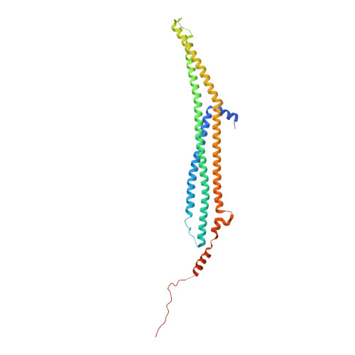
4WPC

Crystal structure of Rgd1p F-BAR domain in complex with inositol phosphate


Experimental Data Snapshot

  • Method: X-RAY DIFFRACTION
  • Resolution: 3.34 Å
  • R-Value Free: 0.300 
  • R-Value Work: 0.261 
  • R-Value Observed: 0.263 

wwPDB Validation   3D Report Full Report


Ligand Structure Quality Assessment 


This is version 1.5 of the entry. See complete history


Literature

Comparison of Saccharomyces cerevisiae F-BAR Domain Structures Reveals a Conserved Inositol Phosphate Binding Site.

Moravcevic, K.Alvarado, D.Schmitz, K.R.Kenniston, J.A.Mendrola, J.M.Ferguson, K.M.Lemmon, M.A.

(2015) Structure 23: 352-363

  • DOI: https://doi.org/10.1016/j.str.2014.12.009
  • Primary Citation of Related Structures:  
    4WPC, 4WPE

  • PubMed Abstract: 

    F-BAR domains control membrane interactions in endocytosis, cytokinesis, and cell signaling. Although they are generally thought to bind curved membranes containing negatively charged phospholipids, numerous functional studies argue that differences in lipid-binding selectivities of F-BAR domains are functionally important. Here, we compare membrane-binding properties of the Saccharomyces cerevisiae F-BAR domains in vitro and in vivo. Whereas some F-BAR domains (such as Bzz1p and Hof1p F-BARs) bind equally well to all phospholipids, the F-BAR domain from the RhoGAP Rgd1p preferentially binds phosphoinositides. We determined X-ray crystal structures of F-BAR domains from Hof1p and Rgd1p, the latter bound to an inositol phosphate. The structures explain phospholipid-binding selectivity differences and reveal an F-BAR phosphoinositide binding site that is fully conserved in a mammalian RhoGAP called Gmip and is partly retained in certain other F-BAR domains. Our findings reveal previously unappreciated determinants of F-BAR domain lipid-binding specificity and provide a basis for its prediction from sequence.


  • Organizational Affiliation

    Department of Biochemistry and Biophysics, University of Pennsylvania Perelman School of Medicine, Philadelphia, PA 19014, USA; Graduate Group in Biochemistry and Molecular Biophysics, University of Pennsylvania Perelman School of Medicine, Philadelphia, PA 19014, USA.


Macromolecules
Find similar proteins by:  (by identity cutoff)  |  3D Structure
Entity ID: 1
MoleculeChains Sequence LengthOrganismDetailsImage
RHO GTPase-activating protein RGD1
A, B
317Saccharomyces cerevisiae S288CMutation(s): 0 
Gene Names: RGD1YBR260CYBR1728
UniProt
Find proteins for P38339 (Saccharomyces cerevisiae (strain ATCC 204508 / S288c))
Explore P38339 
Go to UniProtKB:  P38339
Entity Groups  
Sequence Clusters30% Identity50% Identity70% Identity90% Identity95% Identity100% Identity
UniProt GroupP38339
Sequence Annotations
Expand
  • Reference Sequence
Small Molecules
Experimental Data & Validation

Experimental Data

  • Method: X-RAY DIFFRACTION
  • Resolution: 3.34 Å
  • R-Value Free: 0.300 
  • R-Value Work: 0.261 
  • R-Value Observed: 0.263 
  • Space Group: C 1 2 1
Unit Cell:
Length ( Å )Angle ( ˚ )
a = 178.993α = 90
b = 74.11β = 104.27
c = 105.734γ = 90
Software Package:
Software NamePurpose
PHENIXrefinement
PHASERphasing
HKL-2000data scaling

Structure Validation

View Full Validation Report


Ligand Structure Quality Assessment 


Entry History 

Deposition Data

Revision History  (Full details and data files)

  • Version 1.0: 2014-12-24
    Type: Initial release
  • Version 1.1: 2015-02-11
    Changes: Database references
  • Version 1.2: 2015-02-18
    Changes: Database references
  • Version 1.3: 2015-09-23
    Changes: Non-polymer description
  • Version 1.4: 2020-10-14
    Changes: Derived calculations, Structure summary
  • Version 1.5: 2023-09-27
    Changes: Data collection, Database references, Refinement description