4WMO

Selenomethionine derivative of Xenopus laevis embryonic epidermal lectin carbohydrate-binding domain


Experimental Data Snapshot

  • Method: X-RAY DIFFRACTION
  • Resolution: 2.30 Å
  • R-Value Free: 0.188 
  • R-Value Work: 0.160 
  • R-Value Observed: 0.161 

wwPDB Validation   3D Report Full Report


This is version 1.3 of the entry. See complete history


Literature

Structures of Xenopus Embryonic Epidermal Lectin Reveal a Conserved Mechanism of Microbial Glycan Recognition.

Wangkanont, K.Wesener, D.A.Vidani, J.A.Kiessling, L.L.Forest, K.T.

(2016) J Biol Chem 291: 5596-5610

  • DOI: https://doi.org/10.1074/jbc.M115.709212
  • Primary Citation of Related Structures:  
    4WMO, 4WN0

  • PubMed Abstract: 

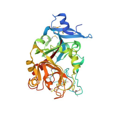
    Intelectins (X-type lectins), broadly distributed throughout chordates, have been implicated in innate immunity. Xenopus laevis embryonic epidermal lectin (XEEL), an intelectin secreted into environmental water by the X. laevis embryo, is postulated to function as a defense against microbes. XEEL is homologous (64% identical) to human intelectin-1 (hIntL-1), which is also implicated in innate immune defense. We showed previously that hIntL-1 binds microbial glycans bearing exocyclic vicinal diol groups. It is unknown whether XEEL has the same ligand specificity. Also unclear is whether XEEL and hIntL-1 have similar quaternary structures, as XEEL lacks the corresponding cysteine residues in hIntL-1 that stabilize the disulfide-linked trimer. These observations prompted us to further characterize XEEL. We found that hIntL-1 and XEEL have similar structural features. Even without the corresponding intermolecular disulfide bonds present in hIntL-1, the carbohydrate recognition domain of XEEL (XEELCRD) forms a stable trimer in solution. The structure of XEELCRD in complex with d-glycerol-1-phosphate, a residue present in microbe-specific glycans, indicated that the exocyclic vicinal diol coordinates to a protein-bound calcium ion. This ligand-binding mode is conserved between XEEL and hIntL-1. The domain architecture of full-length XEEL is reminiscent of a barbell, with two sets of three glycan-binding sites oriented in opposite directions. This orientation is consistent with our observation that XEEL can promote the agglutination of specific serotypes of Streptococcus pneumoniae. These data support a role for XEEL in innate immunity, and they highlight structural and functional conservation of X-type lectins among chordates.


  • Organizational Affiliation

    From the Departments of Chemistry.


Macromolecules
Find similar proteins by:  (by identity cutoff)  |  3D Structure
Entity ID: 1
MoleculeChains Sequence LengthOrganismDetailsImage
XEEL protein289Xenopus laevisMutation(s): 0 
Gene Names: XEEL
UniProt
Find proteins for Q5PPM0 (Xenopus laevis)
Explore Q5PPM0 
Go to UniProtKB:  Q5PPM0
Entity Groups  
Sequence Clusters30% Identity50% Identity70% Identity90% Identity95% Identity100% Identity
UniProt GroupQ5PPM0
Sequence Annotations
Expand
  • Reference Sequence
Small Molecules
Ligands 2 Unique
IDChains Name / Formula / InChI Key2D Diagram3D Interactions
1PE
Query on 1PE

Download Ideal Coordinates CCD File 
AA [auth D]
EA [auth E]
FA [auth E]
J [auth F]
JA [auth A]
AA [auth D],
EA [auth E],
FA [auth E],
J [auth F],
JA [auth A],
K [auth F],
KA [auth A],
LA [auth A],
O [auth C],
P [auth C],
Q [auth C],
U [auth B],
V [auth B],
Z [auth D]
PENTAETHYLENE GLYCOL
C10 H22 O6
JLFNLZLINWHATN-UHFFFAOYSA-N
CA
Query on CA

Download Ideal Coordinates CCD File 
BA [auth E]
CA [auth E]
DA [auth E]
G [auth F]
GA [auth A]
BA [auth E],
CA [auth E],
DA [auth E],
G [auth F],
GA [auth A],
H [auth F],
HA [auth A],
I [auth F],
IA [auth A],
L [auth C],
M [auth C],
N [auth C],
R [auth B],
S [auth B],
T [auth B],
W [auth D],
X [auth D],
Y [auth D]
CALCIUM ION
Ca
BHPQYMZQTOCNFJ-UHFFFAOYSA-N
Modified Residues  1 Unique
IDChains TypeFormula2D DiagramParent
MSE
Query on MSE
A [auth F]
B [auth C]
C [auth B]
D
E
A [auth F],
B [auth C],
C [auth B],
D,
E,
F [auth A]
L-PEPTIDE LINKINGC5 H11 N O2 SeMET
Experimental Data & Validation

Experimental Data

  • Method: X-RAY DIFFRACTION
  • Resolution: 2.30 Å
  • R-Value Free: 0.188 
  • R-Value Work: 0.160 
  • R-Value Observed: 0.161 
  • Space Group: P 1 21 1
Unit Cell:
Length ( Å )Angle ( ˚ )
a = 123.654α = 90
b = 111.069β = 119.72
c = 123.611γ = 90
Software Package:
Software NamePurpose
SCALEPACKdata scaling
PHASERphasing
PHENIXrefinement
PDB_EXTRACTdata extraction

Structure Validation

View Full Validation Report


Entry History 

Deposition Data

Revision History  (Full details and data files)

  • Version 1.0: 2016-01-20
    Type: Initial release
  • Version 1.1: 2016-03-23
    Changes: Database references
  • Version 1.2: 2017-11-22
    Changes: Database references, Derived calculations, Refinement description
  • Version 1.3: 2023-12-27
    Changes: Data collection, Database references, Derived calculations