4WH9

Structure of the CDC25B Phosphatase Catalytic Domain with Bound Inhibitor


Experimental Data Snapshot

  • Method: X-RAY DIFFRACTION
  • Resolution: 1.50 Å
  • R-Value Free: 0.152 
  • R-Value Work: 0.126 
  • R-Value Observed: 0.127 

wwPDB Validation   3D Report Full Report


Ligand Structure Quality Assessment 


This is version 1.5 of the entry. See complete history


Literature

Inhibition of CDC25B Phosphatase Through Disruption of Protein-Protein Interaction.

Lund, G.Dudkin, S.Borkin, D.Ni, W.Grembecka, J.Cierpicki, T.

(2015) ACS Chem Biol 10: 390-394

  • DOI: https://doi.org/10.1021/cb500883h
  • Primary Citation of Related Structures:  
    4WH7, 4WH9

  • PubMed Abstract: 

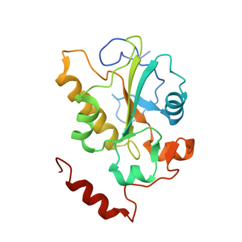
    CDC25 phosphatases are key cell cycle regulators and represent very attractive but challenging targets for anticancer drug discovery. Here, we explored whether fragment-based screening represents a valid approach to identify inhibitors of CDC25B. This resulted in identification of 2-fluoro-4-hydroxybenzonitrile, which directly binds to the catalytic domain of CDC25B. Interestingly, NMR data and the crystal structure demonstrate that this compound binds to the pocket distant from the active site and adjacent to the protein-protein interaction interface with CDK2/Cyclin A substrate. Furthermore, we developed a more potent analogue that disrupts CDC25B interaction with CDK2/Cyclin A and inhibits dephosphorylation of CDK2. Based on these studies, we provide a proof of concept that targeting CDC25 phosphatases by inhibiting their protein-protein interactions with CDK2/Cyclin A substrate represents a novel, viable opportunity to target this important class of enzymes.


  • Organizational Affiliation

    Department of Pathology, University of Michigan , 4510C MSRBI 1150 West Medical Center Drive, Ann Arbor, Michigan 48109-5620, United States.


Macromolecules
Find similar proteins by:  (by identity cutoff)  |  3D Structure
Entity ID: 1
MoleculeChains Sequence LengthOrganismDetailsImage
M-phase inducer phosphatase 2183Homo sapiensMutation(s): 1 
Gene Names: CDC25BCDC25HU2
EC: 3.1.3.48
UniProt & NIH Common Fund Data Resources
Find proteins for P30305 (Homo sapiens)
Explore P30305 
Go to UniProtKB:  P30305
PHAROS:  P30305
GTEx:  ENSG00000101224 
Entity Groups  
Sequence Clusters30% Identity50% Identity70% Identity90% Identity95% Identity100% Identity
UniProt GroupP30305
Sequence Annotations
Expand
  • Reference Sequence
Experimental Data & Validation

Experimental Data

  • Method: X-RAY DIFFRACTION
  • Resolution: 1.50 Å
  • R-Value Free: 0.152 
  • R-Value Work: 0.126 
  • R-Value Observed: 0.127 
  • Space Group: P 21 21 21
Unit Cell:
Length ( Å )Angle ( ˚ )
a = 51.66α = 90
b = 71.47β = 90
c = 73.94γ = 90
Software Package:
Software NamePurpose
REFMACrefinement
iMOSFLMdata reduction
SCALAdata scaling
MOLREPphasing

Structure Validation

View Full Validation Report


Ligand Structure Quality Assessment 


Entry History & Funding Information

Deposition Data


Funding OrganizationLocationGrant Number
National Institutes of Health/National Cancer Institute (NIH/NCI)United StatesCA181185

Revision History  (Full details and data files)

  • Version 1.0: 2014-12-10
    Type: Initial release
  • Version 1.1: 2015-02-04
    Changes: Derived calculations
  • Version 1.2: 2015-03-04
    Changes: Database references
  • Version 1.3: 2017-09-27
    Changes: Author supporting evidence, Derived calculations, Source and taxonomy
  • Version 1.4: 2019-12-04
    Changes: Author supporting evidence
  • Version 1.5: 2023-09-27
    Changes: Data collection, Database references, Refinement description