4WH3

N-acetylhexosamine 1-kinase in complex with ATP


Experimental Data Snapshot

  • Method: X-RAY DIFFRACTION
  • Resolution: 1.80 Å
  • R-Value Free: 0.222 
  • R-Value Work: 0.189 
  • R-Value Observed: 0.191 

wwPDB Validation   3D Report Full Report


Ligand Structure Quality Assessment 


This is version 1.2 of the entry. See complete history


Literature

Open-close structural change upon ligand binding and two magnesium ions required for the catalysis of N-acetylhexosamine 1-kinase

Sato, M.Arakawa, T.Nam, Y.W.Nishimoto, M.Kitaoka, M.Fushinobu, S.

(2015) Biochim Biophys Acta 1854: 333-340

  • DOI: https://doi.org/10.1016/j.bbapap.2015.01.011
  • Primary Citation of Related Structures:  
    4WH1, 4WH2, 4WH3

  • PubMed Abstract: 

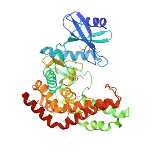
    Infant gut-associated bifidobacteria possess a metabolic pathway to utilize lacto-N-biose (Gal-β1,3-GlcNAc) and galacto-N-biose (Gal-β1,3-GalNAc) from human milk and glycoconjugates specifically. In this pathway, N-acetylhexosamine 1-kinase (NahK) catalyzes the phosphorylation of GlcNAc or GalNAc at the anomeric C1 position with ATP. Crystal structures of NahK have only been determined in the closed state. In this study, we determined open state structures of NahK in three different forms (apo, ADP complex, and ATP complex). A comparison of the open and closed state structures revealed an induced fit structural change defined by two rigid domains. ATP binds to the small N-terminal domain, and binding of the N-acetylhexosamine substrate to the large C-terminal domain induces a closing conformational change with a rotation angle of 16°. In the nucleotide binding site, two magnesium ions bridging the α-γ and β-γ phosphates were identified. A mutational analysis indicated that a residue coordinating both of the two magnesium ions (Asp228) is essential for catalysis. The involvement of two magnesium ions in the catalytic machinery is structurally similar to the catalytic structures of protein kinases and aminoglycoside phosphotransferases, but distinct from the structures of other anomeric kinases or sugar 6-kinases. These findings help to elucidate the possible evolutionary adaptation of substrate specificities and induced fit mechanism.


  • Organizational Affiliation

    Department of Biotechnology, The University of Tokyo, Tokyo 113-8657, Japan.


Macromolecules
Find similar proteins by:  (by identity cutoff)  |  3D Structure
Entity ID: 1
MoleculeChains Sequence LengthOrganismDetailsImage
N-acetylhexosamine 1-kinase367Bifidobacterium longum subsp. longum JCM 1217Mutation(s): 1 
Gene Names: nahKlnpBBLLJ_1622
EC: 2.7.1.162
UniProt
Find proteins for E8MF12 (Bifidobacterium longum subsp. longum (strain ATCC 15707 / DSM 20219 / JCM 1217 / NCTC 11818 / E194b))
Explore E8MF12 
Go to UniProtKB:  E8MF12
Entity Groups  
Sequence Clusters30% Identity50% Identity70% Identity90% Identity95% Identity100% Identity
UniProt GroupE8MF12
Sequence Annotations
Expand
  • Reference Sequence
Small Molecules
Ligands 4 Unique
IDChains Name / Formula / InChI Key2D Diagram3D Interactions
ATP
Query on ATP

Download Ideal Coordinates CCD File 
B [auth A]ADENOSINE-5'-TRIPHOSPHATE
C10 H16 N5 O13 P3
ZKHQWZAMYRWXGA-KQYNXXCUSA-N
GOL
Query on GOL

Download Ideal Coordinates CCD File 
E [auth A],
F [auth A],
G [auth A],
H [auth A],
I [auth A]
GLYCEROL
C3 H8 O3
PEDCQBHIVMGVHV-UHFFFAOYSA-N
ACY
Query on ACY

Download Ideal Coordinates CCD File 
J [auth A],
K [auth A]
ACETIC ACID
C2 H4 O2
QTBSBXVTEAMEQO-UHFFFAOYSA-N
MG
Query on MG

Download Ideal Coordinates CCD File 
C [auth A],
D [auth A]
MAGNESIUM ION
Mg
JLVVSXFLKOJNIY-UHFFFAOYSA-N
Modified Residues  1 Unique
IDChains TypeFormula2D DiagramParent
MSE
Query on MSE
A
L-PEPTIDE LINKINGC5 H11 N O2 SeMET
Experimental Data & Validation

Experimental Data

  • Method: X-RAY DIFFRACTION
  • Resolution: 1.80 Å
  • R-Value Free: 0.222 
  • R-Value Work: 0.189 
  • R-Value Observed: 0.191 
  • Space Group: P 21 21 21
Unit Cell:
Length ( Å )Angle ( ˚ )
a = 44.03α = 90
b = 88.471β = 90
c = 105.219γ = 90
Software Package:
Software NamePurpose
HKL-2000data processing
PHENIXphasing
PHENIXmodel building
PHENIXrefinement
Cootrefinement

Structure Validation

View Full Validation Report


Ligand Structure Quality Assessment 


Entry History & Funding Information

Deposition Data


Funding OrganizationLocationGrant Number
Ministry of Agriculture, Forestry and FisheriesJapan--

Revision History  (Full details and data files)

  • Version 1.0: 2015-02-18
    Type: Initial release
  • Version 1.1: 2015-02-25
    Changes: Database references
  • Version 1.2: 2020-02-05
    Changes: Data collection, Database references, Derived calculations, Other, Source and taxonomy