4WCX

Crystal structure of HydG: A maturase of the [FeFe]-hydrogenase


Experimental Data Snapshot

  • Method: X-RAY DIFFRACTION
  • Resolution: 1.59 Å
  • R-Value Free: 0.213 
  • R-Value Work: 0.180 
  • R-Value Observed: 0.180 

wwPDB Validation   3D Report Full Report


Ligand Structure Quality Assessment 


This is version 1.1 of the entry. See complete history


Literature

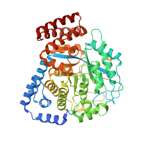
X-ray crystallographic and EPR spectroscopic analysis of HydG, a maturase in [FeFe]-hydrogenase H-cluster assembly.

Dinis, P.Suess, D.L.Fox, S.J.Harmer, J.E.Driesener, R.C.De La Paz, L.Swartz, J.R.Essex, J.W.Britt, R.D.Roach, P.L.

(2015) Proc Natl Acad Sci U S A 112: 1362-1367

  • DOI: https://doi.org/10.1073/pnas.1417252112
  • Primary Citation of Related Structures:  
    4WCX

  • PubMed Abstract: 

    Hydrogenases use complex metal cofactors to catalyze the reversible formation of hydrogen. In [FeFe]-hydrogenases, the H-cluster cofactor includes a diiron subcluster containing azadithiolate, three CO, and two CN(-) ligands. During the assembly of the H cluster, the radical S-adenosyl methionine (SAM) enzyme HydG lyses the substrate tyrosine to yield the diatomic ligands. These diatomic products form an enzyme-bound Fe(CO)x(CN)y synthon that serves as a precursor for eventual H-cluster assembly. To further elucidate the mechanism of this complex reaction, we report the crystal structure and EPR analysis of HydG. At one end of the HydG (βα)8 triosephosphate isomerase (TIM) barrel, a canonical [4Fe-4S] cluster binds SAM in close proximity to the proposed tyrosine binding site. At the opposite end of the active-site cavity, the structure reveals the auxiliary Fe-S cluster in two states: one monomer contains a [4Fe-5S] cluster, and the other monomer contains a [5Fe-5S] cluster consisting of a [4Fe-4S] cubane bridged by a μ2-sulfide ion to a mononuclear Fe(2+) center. This fifth iron is held in place by a single highly conserved protein-derived ligand: histidine 265. EPR analysis confirms the presence of the [5Fe-5S] cluster, which on incubation with cyanide, undergoes loss of the labile iron to yield a [4Fe-4S] cluster. We hypothesize that the labile iron of the [5Fe-5S] cluster is the site of Fe(CO)x(CN)y synthon formation and that the limited bonding between this iron and HydG may facilitate transfer of the intact synthon to its cognate acceptor for subsequent H-cluster assembly.


  • Organizational Affiliation

    Chemistry and the Institute for Life Sciences, University of Southampton, Southampton SO17 1BJ, United Kingdom;


Macromolecules
Find similar proteins by:  (by identity cutoff)  |  3D Structure
Entity ID: 1
MoleculeChains Sequence LengthOrganismDetailsImage
Biotin and thiamin synthesis associatedA,
B [auth C]
480Thermoanaerobacter italicus Ab9Mutation(s): 0 
Gene Names: Thit_0582
UniProt
Find proteins for D3T7F1 (Thermoanaerobacter italicus (strain DSM 9252 / Ab9))
Explore D3T7F1 
Go to UniProtKB:  D3T7F1
Entity Groups  
Sequence Clusters30% Identity50% Identity70% Identity90% Identity95% Identity100% Identity
UniProt GroupD3T7F1
Sequence Annotations
Expand
  • Reference Sequence
Small Molecules
Ligands 7 Unique
IDChains Name / Formula / InChI Key2D Diagram3D Interactions
SAM
Query on SAM

Download Ideal Coordinates CCD File 
L [auth C]S-ADENOSYLMETHIONINE
C15 H22 N6 O5 S
MEFKEPWMEQBLKI-FCKMPRQPSA-N
SF4
Query on SF4

Download Ideal Coordinates CCD File 
C [auth A],
D [auth A],
J [auth C],
K [auth C]
IRON/SULFUR CLUSTER
Fe4 S4
LJBDFODJNLIPKO-UHFFFAOYSA-N
MET
Query on MET

Download Ideal Coordinates CCD File 
E [auth A]METHIONINE
C5 H11 N O2 S
FFEARJCKVFRZRR-BYPYZUCNSA-N
ALA
Query on ALA

Download Ideal Coordinates CCD File 
G [auth A]ALANINE
C3 H7 N O2
QNAYBMKLOCPYGJ-REOHCLBHSA-N
FE
Query on FE

Download Ideal Coordinates CCD File 
F [auth A]FE (III) ION
Fe
VTLYFUHAOXGGBS-UHFFFAOYSA-N
H2S
Query on H2S

Download Ideal Coordinates CCD File 
H [auth A],
M [auth C]
HYDROSULFURIC ACID
H2 S
RWSOTUBLDIXVET-UHFFFAOYSA-N
NA
Query on NA

Download Ideal Coordinates CCD File 
I [auth A],
N [auth C],
O [auth C]
SODIUM ION
Na
FKNQFGJONOIPTF-UHFFFAOYSA-N
Experimental Data & Validation

Experimental Data

  • Method: X-RAY DIFFRACTION
  • Resolution: 1.59 Å
  • R-Value Free: 0.213 
  • R-Value Work: 0.180 
  • R-Value Observed: 0.180 
  • Space Group: P 1
Unit Cell:
Length ( Å )Angle ( ˚ )
a = 54.13α = 89.59
b = 56.19β = 83.62
c = 84.92γ = 66.84
Software Package:
Software NamePurpose
PHENIXrefinement
XDSdata reduction
SCALAdata scaling
PHENIXphasing

Structure Validation

View Full Validation Report


Ligand Structure Quality Assessment 


Entry History 

Deposition Data

Revision History  (Full details and data files)

  • Version 1.0: 2015-02-04
    Type: Initial release
  • Version 1.1: 2015-02-11
    Changes: Database references