4WAE

Phosphatidylinositol 4-kinase III beta crystallized with ATP


Experimental Data Snapshot

  • Method: X-RAY DIFFRACTION
  • Resolution: 3.32 Å
  • R-Value Free: 0.244 
  • R-Value Work: 0.182 
  • R-Value Observed: 0.185 

wwPDB Validation   3D Report Full Report


This is version 1.3 of the entry. See complete history


Literature

Highly Selective Phosphatidylinositol 4-Kinase III beta Inhibitors and Structural Insight into Their Mode of Action.

Mejdrova, I.Chalupska, D.Kogler, M.Sala, M.Plackova, P.Baumlova, A.Hrebabecky, H.Prochazkova, E.Dejmek, M.Guillon, R.Strunin, D.Weber, J.Lee, G.Birkus, G.Mertlikova-Kaiserova, H.Boura, E.Nencka, R.

(2015) J Med Chem 58: 3767-3793

  • DOI: https://doi.org/10.1021/acs.jmedchem.5b00499
  • Primary Citation of Related Structures:  
    4WAE, 4WAG

  • PubMed Abstract: 

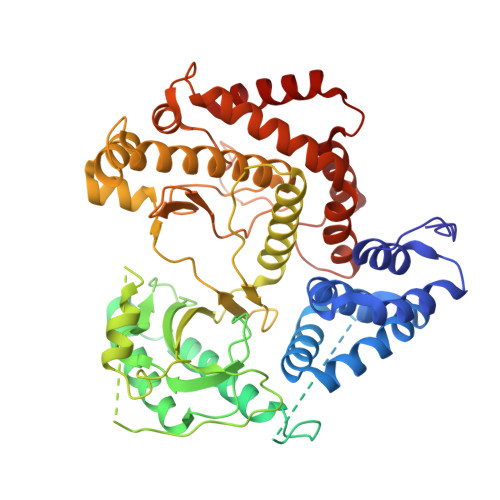
    Phosphatidylinositol 4-kinase IIIβ is a cellular lipid kinase pivotal to pathogenesis of various RNA viruses. These viruses hijack the enzyme in order to modify the structure of intracellular membranes and use them for the construction of functional replication machinery. Selective inhibitors of this enzyme are potential broad-spectrum antiviral agents, as inhibition of this enzyme results in the arrest of replication of PI4K IIIβ-dependent viruses. Herein, we report a detailed study of novel selective inhibitors of PI4K IIIβ, which exert antiviral activity against a panel of single-stranded positive-sense RNA viruses. Our crystallographic data show that the inhibitors occupy the binding site for the adenine ring of the ATP molecule and therefore prevent the phosphorylation reaction.


  • Organizational Affiliation

    ‡Gilead Sciences, Inc., 333 Lakeside Drive, Foster City, California 94404, United States.


Macromolecules
Find similar proteins by:  (by identity cutoff)  |  3D Structure
Entity ID: 1
MoleculeChains Sequence LengthOrganismDetailsImage
Phosphatidylinositol 4-kinase beta,Phosphatidylinositol 4-kinase beta572Homo sapiensMutation(s): 0 
Gene Names: PI4KBPIK4CB
EC: 2.7.1.67
UniProt & NIH Common Fund Data Resources
Find proteins for Q9UBF8 (Homo sapiens)
Explore Q9UBF8 
Go to UniProtKB:  Q9UBF8
PHAROS:  Q9UBF8
GTEx:  ENSG00000143393 
Entity Groups  
Sequence Clusters30% Identity50% Identity70% Identity90% Identity95% Identity100% Identity
UniProt GroupQ9UBF8
Sequence Annotations
Expand
  • Reference Sequence
Small Molecules
Ligands 1 Unique
IDChains Name / Formula / InChI Key2D Diagram3D Interactions
ATP
Query on ATP

Download Ideal Coordinates CCD File 
B [auth A]ADENOSINE-5'-TRIPHOSPHATE
C10 H16 N5 O13 P3
ZKHQWZAMYRWXGA-KQYNXXCUSA-N
Experimental Data & Validation

Experimental Data

  • Method: X-RAY DIFFRACTION
  • Resolution: 3.32 Å
  • R-Value Free: 0.244 
  • R-Value Work: 0.182 
  • R-Value Observed: 0.185 
  • Space Group: P 43
Unit Cell:
Length ( Å )Angle ( ˚ )
a = 108.327α = 90
b = 108.327β = 90
c = 55.189γ = 90
Software Package:
Software NamePurpose
PHENIXrefinement
XDSdata reduction
XSCALEdata scaling
PHASERphasing

Structure Validation

View Full Validation Report


Entry History & Funding Information

Deposition Data


Funding OrganizationLocationGrant Number
Ministry of Education, Youth and Sports of the Czech RepublicCzech RepublicLO1302

Revision History  (Full details and data files)

  • Version 1.0: 2015-05-20
    Type: Initial release
  • Version 1.1: 2015-05-27
    Changes: Database references
  • Version 1.2: 2017-09-06
    Changes: Author supporting evidence
  • Version 1.3: 2024-01-10
    Changes: Data collection, Database references, Refinement description