


Crystal structure of the periplasmic domain of subunit II of cytochrome oxidase (CoxB) of Bradyrhizobium japonicum
Quade, N., Abicht, H.K., Hennecke, H., Glockshuber, R.To be published.
Experimental Data Snapshot
wwPDB Validation 3D Report Full Report

Entity ID: 1 | |||||
|---|---|---|---|---|---|
| Molecule | Chains | Sequence Length | Organism | Details | Image |
| Cytochrome c oxidase subunit 2 | 139 | Bradyrhizobium diazoefficiens USDA 110 | Mutation(s): 0 Gene Names: coxB, blr1170 EC: 1.9.3.1 | 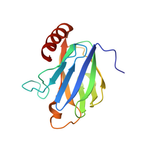 | |
UniProt | |||||
Find proteins for H7C6E5 (Bradyrhizobium diazoefficiens (strain JCM 10833 / BCRC 13528 / IAM 13628 / NBRC 14792 / USDA 110)) Explore H7C6E5 Go to UniProtKB: H7C6E5 | |||||
Entity Groups | |||||
| Sequence Clusters | 30% Identity50% Identity70% Identity90% Identity95% Identity100% Identity | ||||
| UniProt Group | H7C6E5 | ||||
Sequence AnnotationsExpand | |||||
| |||||
| Ligands 1 Unique | |||||
|---|---|---|---|---|---|
| ID | Chains | Name / Formula / InChI Key | 2D Diagram | 3D Interactions | |
| CU Query on CU | B [auth A], C [auth A] | COPPER (II) ION Cu JPVYNHNXODAKFH-UHFFFAOYSA-N |  | ||
| Length ( Å ) | Angle ( ˚ ) |
|---|---|
| a = 32.431 | α = 90 |
| b = 38.612 | β = 97.63 |
| c = 59.644 | γ = 90 |
| Software Name | Purpose |
|---|---|
| REFMAC | refinement |
| XDS | data reduction |
| Coot | model building |
| PDB_EXTRACT | data extraction |
| XSCALE | data scaling |
| PHASER | phasing |
| XSCALE | data reduction |
RCSB PDB (citation) is hosted by
RCSB PDB is a member of the

