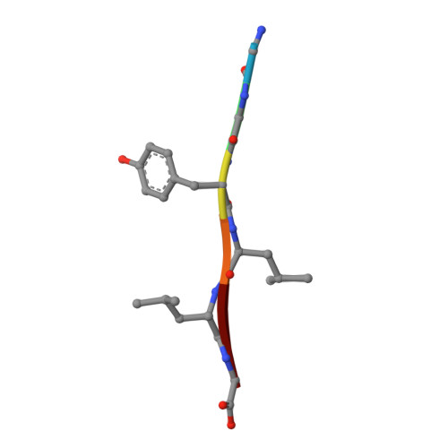
Crystal Structures of Polymorphic Prion Protein beta 1 Peptides Reveal Variable Steric Zipper Conformations.
Yu, L., Lee, S.J., Yee, V.C.(2015) Biochemistry 54: 3640-3648
- PubMed: 25978088
- DOI: https://doi.org/10.1021/acs.biochem.5b00425
- Primary Citation of Related Structures:
4TUT, 4UBY, 4UBZ, 4W5L, 4W5M, 4W5P, 4W5Y, 4W67, 4W71, 4WBU, 4WBV - PubMed Abstract:
The pathogenesis of prion diseases is associated with the conformational conversion of normal, predominantly α-helical prion protein (PrP(C)) into a pathogenic form that is enriched with β-sheets (PrP(Sc)). Several PrP(C) crystal structures have revealed β1-mediated intermolecular sheets, suggesting that the β1 strand may contribute to a possible initiation site for β-sheet-mediated PrP(Sc) propagation. This β1 strand contains the polymorphic residue 129 that influences disease susceptibility and phenotype. To investigate the effect of the residue 129 polymorphism on the conformation of amyloid-like continuous β-sheets formed by β1, crystal structures of β1 peptides containing each of the polymorphic residues were determined. To probe the conformational influence of the peptide construct design, four different lengths of β1 peptides were studied. From the 12 peptides studied, 11 yielded crystal structures ranging in resolution from 0.9 to 1.4 Å. This ensemble of β1 crystal structures reveals conformational differences that are influenced by both the nature of the polymorphic residue and the extent of the peptide construct, indicating that comprehensive studies in which peptide constructs vary are a more rigorous approach to surveying conformational possibilities.
Organizational Affiliation:
Department of Biochemistry, Case Western Reserve University, Cleveland, Ohio 44106, United States.


