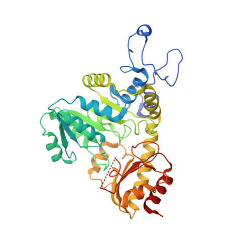
4U2H

The crystal structure of apo CalE6, a methionine gamma lyase from Micromonospora echinospora


Experimental Data Snapshot

  • Method: X-RAY DIFFRACTION
  • Resolution: 1.85 Å
  • R-Value Free: 0.232 
  • R-Value Work: 0.209 

wwPDB Validation   3D Report Full Report


This is version 1.1 of the entry. See complete history


Literature

Identification and Characterization of a Methionine gamma-Lyase in the Calicheamicin Biosynthetic Cluster of Micromonospora echinospora

Song, H.Xu, R.Guo, Z.

(2015) Chembiochem 16: 100-109

  • DOI: https://doi.org/10.1002/cbic.201402489
  • Primary Citation of Related Structures:  
    4U1T, 4U2H

  • PubMed Abstract: 

    CalE6 is a previously uncharacterized protein involved in the biosynthesis of calicheamicins in Micromonospora echinospora. It is a pyridoxal-5'-phosphate-dependent enzyme and exhibits high sequence homology to cystathionine γ-lyases and cystathionine γ-synthases. However, it was found to be active towards methionine and to convert this amino acid into α-ketobutyrate, ammonium, and methanethiol. The crystal structure of the cofactor-bound holoenzyme was resolved at 2.0 Å; it contains two active site residues, Gly105 and Val322, specific for methionine γ-lyases. Modeling of methionine into the active site allows identification of the active site residues responsible for substrate recognition and catalysis. These findings support that CalE6 is a putative methionine γ-lyase producing methanethiol as a building block in biosynthesis of calicheamicins.


  • Organizational Affiliation

    Department of Chemistry and State Key Laboratory of Molecular Neuroscience, The Hong Kong University of Science and Technology, Clear Water Bay, Kowloon (Hong Kong).


Macromolecules
Find similar proteins by:  (by identity cutoff)  |  3D Structure
Entity ID: 1
MoleculeChains Sequence LengthOrganismDetailsImage
CalE6
A, B, C, D, E
A, B, C, D, E, F, G, H
389Micromonospora echinosporaMutation(s): 0 
Gene Names: calE6
EC: 4.4.1.11
UniProt
Find proteins for Q8KNG3 (Micromonospora echinospora)
Explore Q8KNG3 
Go to UniProtKB:  Q8KNG3
Entity Groups  
Sequence Clusters30% Identity50% Identity70% Identity90% Identity95% Identity100% Identity
UniProt GroupQ8KNG3
Sequence Annotations
Expand
  • Reference Sequence
Small Molecules
Ligands 1 Unique
IDChains Name / Formula / InChI Key2D Diagram3D Interactions
SO4
Query on SO4

Download Ideal Coordinates CCD File 
I [auth A]
J [auth A]
K [auth B]
L [auth B]
M [auth C]
I [auth A],
J [auth A],
K [auth B],
L [auth B],
M [auth C],
N [auth C],
O [auth D],
P [auth D],
Q [auth E],
R [auth E],
S [auth F],
T [auth F],
U [auth G],
V [auth G],
W [auth G],
X [auth H]
SULFATE ION
O4 S
QAOWNCQODCNURD-UHFFFAOYSA-L
Experimental Data & Validation

Experimental Data

  • Method: X-RAY DIFFRACTION
  • Resolution: 1.85 Å
  • R-Value Free: 0.232 
  • R-Value Work: 0.209 
  • Space Group: I 2 2 2
Unit Cell:
Length ( Å )Angle ( ˚ )
a = 145.204α = 90
b = 145.395β = 90
c = 346.119γ = 90
Software Package:
Software NamePurpose
HKL-2000data collection
HKL-2000data processing
HKL-2000data scaling
PHENIXrefinement
PHASERphasing
Cootmodel building

Structure Validation

View Full Validation Report


Entry History 

Deposition Data

Revision History  (Full details and data files)

  • Version 1.0: 2015-01-07
    Type: Initial release
  • Version 1.1: 2024-03-20
    Changes: Advisory, Data collection, Database references, Derived calculations, Other, Refinement description, Source and taxonomy