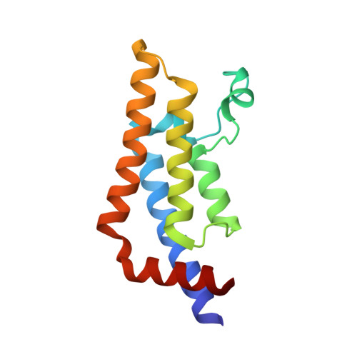
4TU4

Crystal structure of ATAD2A bromodomain complexed with 3-(3,5-dimethyl-1,2-oxazol-4-yl)-5-[(phenylsulfonyl)amino]benzoicacid


Experimental Data Snapshot

  • Method: X-RAY DIFFRACTION
  • Resolution: 1.73 Å
  • R-Value Free: 0.197 
  • R-Value Work: 0.184 
  • R-Value Observed: 0.185 

wwPDB Validation   3D Report Full Report


Ligand Structure Quality Assessment 


This is version 1.4 of the entry. See complete history


Literature

Observed bromodomain flexibility reveals histone peptide- and small molecule ligand-compatible forms of ATAD2.

Poncet-Montange, G.Zhan, Y.Bardenhagen, J.P.Petrocchi, A.Leo, E.Shi, X.Lee, G.R.Leonard, P.G.Geck Do, M.K.Cardozo, M.G.Andersen, J.N.Palmer, W.S.Jones, P.Ladbury, J.E.

(2015) Biochem J 466: 337-346

  • DOI: https://doi.org/10.1042/BJ20140933

  • PubMed Abstract: 

    Preventing histone recognition by bromodomains emerges as an attractive therapeutic approach in cancer. Overexpression of ATAD2 (ATPase family AAA domain-containing 2 isoform A) in cancer cells is associated with poor prognosis making the bromodomain of ATAD2 a promising epigenetic therapeutic target. In the development of an in vitro assay and identification of small molecule ligands, we conducted structure-guided studies which revealed a conformationally flexible ATAD2 bromodomain. Structural studies on apo-, peptide-and small molecule-ATAD2 complexes (by co-crystallization) revealed that the bromodomain adopts a 'closed', histone-compatible conformation and a more 'open' ligand-compatible conformation of the binding site respectively. An unexpected conformational change of the conserved asparagine residue plays an important role in driving the peptide-binding conformation remodelling. We also identified dimethylisoxazole-containing ligands as ATAD2 binders which aided in the validation of the in vitro screen and in the analysis of these conformational studies.


  • Organizational Affiliation

    *Institute for Applied Cancer Science, The University of Texas MD Anderson Cancer Center, Unit 1954, 1515 Holcombe Blvd, Houston, TX 77030, U.S.A.


Macromolecules
Find similar proteins by:  (by identity cutoff)  |  3D Structure
Entity ID: 1
MoleculeChains Sequence LengthOrganismDetailsImage
ATPase family AAA domain-containing protein 2130Homo sapiensMutation(s): 0 
Gene Names: ATAD2L16PRO2000
EC: 3.6.1.3
UniProt & NIH Common Fund Data Resources
Find proteins for Q6PL18 (Homo sapiens)
Explore Q6PL18 
Go to UniProtKB:  Q6PL18
PHAROS:  Q6PL18
GTEx:  ENSG00000156802 
Entity Groups  
Sequence Clusters30% Identity50% Identity70% Identity90% Identity95% Identity100% Identity
UniProt GroupQ6PL18
Sequence Annotations
Expand
  • Reference Sequence
Small Molecules
Ligands 5 Unique
IDChains Name / Formula / InChI Key2D Diagram3D Interactions
37N
Query on 37N

Download Ideal Coordinates CCD File 
E [auth A]3-(3,5-dimethyl-1,2-oxazol-4-yl)-5-[(phenylsulfonyl)amino]benzoic acid
C18 H16 N2 O5 S
FFWKFJNEMDSJKS-UHFFFAOYSA-N
SO4
Query on SO4

Download Ideal Coordinates CCD File 
B [auth A]SULFATE ION
O4 S
QAOWNCQODCNURD-UHFFFAOYSA-L
GOL
Query on GOL

Download Ideal Coordinates CCD File 
C [auth A],
D [auth A]
GLYCEROL
C3 H8 O3
PEDCQBHIVMGVHV-UHFFFAOYSA-N
DMS
Query on DMS

Download Ideal Coordinates CCD File 
G [auth A]DIMETHYL SULFOXIDE
C2 H6 O S
IAZDPXIOMUYVGZ-UHFFFAOYSA-N
CL
Query on CL

Download Ideal Coordinates CCD File 
F [auth A]CHLORIDE ION
Cl
VEXZGXHMUGYJMC-UHFFFAOYSA-M
Binding Affinity Annotations 
IDSourceBinding Affinity
37N Binding MOAD:  4TU4 Kd: 1.75e+5 (nM) from 1 assay(s)
Experimental Data & Validation

Experimental Data

  • Method: X-RAY DIFFRACTION
  • Resolution: 1.73 Å
  • R-Value Free: 0.197 
  • R-Value Work: 0.184 
  • R-Value Observed: 0.185 
  • Space Group: P 65 2 2
Unit Cell:
Length ( Å )Angle ( ˚ )
a = 79.288α = 90
b = 79.288β = 90
c = 137.945γ = 120
Software Package:
Software NamePurpose
REFMACrefinement
SCALAdata scaling
PDB_EXTRACTdata extraction

Structure Validation

View Full Validation Report


Ligand Structure Quality Assessment 


Entry History 

Revision History  (Full details and data files)

  • Version 1.0: 2014-12-24
    Type: Initial release
  • Version 1.1: 2015-02-04
    Changes: Derived calculations
  • Version 1.2: 2015-03-04
    Changes: Database references
  • Version 1.3: 2017-11-22
    Changes: Database references, Derived calculations, Other, Refinement description, Source and taxonomy
  • Version 1.4: 2023-09-27
    Changes: Data collection, Database references, Refinement description