4TPS

Sporulation Inhibitor of DNA Replication, SirA, in complex with Domain I of DnaA


Experimental Data Snapshot

  • Method: X-RAY DIFFRACTION
  • Resolution: 1.65 Å
  • R-Value Free: 0.193 
  • R-Value Work: 0.126 
  • R-Value Observed: 0.130 

wwPDB Validation   3D Report Full Report


This is version 1.2 of the entry. See complete history


Literature

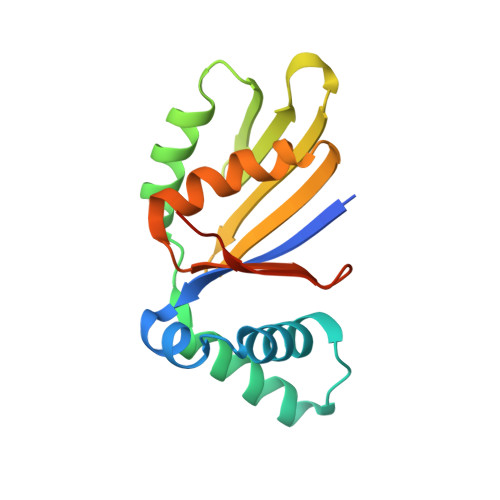
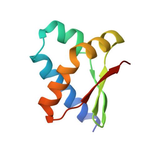
Structure and interactions of the Bacillus subtilis sporulation inhibitor of DNA replication, SirA, with domain I of DnaA.

Jameson, K.H.Rostami, N.Fogg, M.J.Turkenburg, J.P.Grahl, A.Murray, H.Wilkinson, A.J.

(2014) Mol Microbiol 93: 975-991

  • DOI: https://doi.org/10.1111/mmi.12713
  • Primary Citation of Related Structures:  
    4TPS

  • PubMed Abstract: 

    Chromosome copy number in cells is controlled so that the frequency of initiation of DNA replication matches that of cell division. In bacteria, this is achieved through regulation of the interaction between the initiator protein DnaA and specific DNA elements arrayed at the origin of replication. DnaA assembles at the origin and promotes DNA unwinding and the assembly of a replication initiation complex. SirA is a DnaA-interacting protein that inhibits initiation of replication in diploid Bacillus subtilis cells committed to the developmental pathway leading to formation of a dormant spore. Here we present the crystal structure of SirA in complex with the N-terminal domain of DnaA revealing a heterodimeric complex. The interacting surfaces of both proteins are α-helical with predominantly apolar side-chains packing in a hydrophobic interface. Site-directed mutagenesis experiments confirm the importance of this interface for the interaction of the two proteins in vitro and in vivo. Localization of GFP-SirA indicates that the protein accumulates at the replisome in sporulating cells, likely through a direct interaction with DnaA. The SirA interacting surface of DnaA corresponds closely to the HobA-interacting surface of DnaA from Helicobacter pylori even though HobA is an activator of DnaA and SirA is an inhibitor.


  • Organizational Affiliation

    Structural Biology Laboratory, Department of Chemistry, University of York, York, YO10 5DD, UK.


Macromolecules
Find similar proteins by:  (by identity cutoff)  |  3D Structure
Entity ID: 1
MoleculeChains Sequence LengthOrganismDetailsImage
Sporulation inhibitor of replication protein SirA
A, C
154Bacillus subtilis subsp. subtilis str. 168Mutation(s): 0 
Gene Names: sirAyneEyoxFBSU17900
UniProt
Find proteins for P45707 (Bacillus subtilis (strain 168))
Explore P45707 
Go to UniProtKB:  P45707
Entity Groups  
Sequence Clusters30% Identity50% Identity70% Identity90% Identity95% Identity100% Identity
UniProt GroupP45707
Sequence Annotations
Expand
  • Reference Sequence
Find similar proteins by:  (by identity cutoff)  |  3D Structure
Entity ID: 2
MoleculeChains Sequence LengthOrganismDetailsImage
Chromosomal replication initiator protein DnaA
B, D
85Bacillus subtilis subsp. subtilis str. 168Mutation(s): 0 
Gene Names: dnaAdnaHBSU00010
UniProt
Find proteins for P05648 (Bacillus subtilis (strain 168))
Explore P05648 
Go to UniProtKB:  P05648
Entity Groups  
Sequence Clusters30% Identity50% Identity70% Identity90% Identity95% Identity100% Identity
UniProt GroupP05648
Sequence Annotations
Expand
  • Reference Sequence
Experimental Data & Validation

Experimental Data

  • Method: X-RAY DIFFRACTION
  • Resolution: 1.65 Å
  • R-Value Free: 0.193 
  • R-Value Work: 0.126 
  • R-Value Observed: 0.130 
  • Space Group: P 1 21 1
Unit Cell:
Length ( Å )Angle ( ˚ )
a = 77.287α = 90
b = 34.685β = 102.09
c = 84.735γ = 90
Software Package:
Software NamePurpose
REFMACrefinement

Structure Validation

View Full Validation Report


Entry History 

Deposition Data

Revision History  (Full details and data files)

  • Version 1.0: 2014-07-30
    Type: Initial release
  • Version 1.1: 2014-09-10
    Changes: Database references
  • Version 1.2: 2024-04-03
    Changes: Data collection, Database references, Derived calculations, Refinement description