4S1Z

Crystal structure of TRABID NZF1 in complex with K29 linked di-Ubiquitin


Experimental Data Snapshot

  • Method: X-RAY DIFFRACTION
  • Resolution: 3.03 Å
  • R-Value Free: 0.270 
  • R-Value Work: 0.222 
  • R-Value Observed: 0.225 

wwPDB Validation   3D Report Full Report


This is version 1.2 of the entry. See complete history


Literature

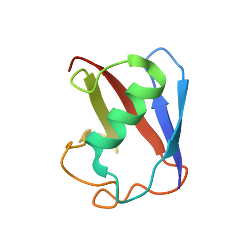
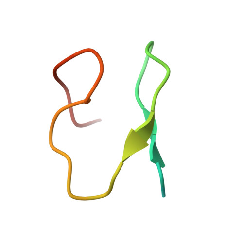
K29-selective ubiquitin binding domain reveals structural basis of specificity and heterotypic nature of k29 polyubiquitin.

Kristariyanto, Y.A.Abdul Rehman, S.A.Campbell, D.G.Morrice, N.A.Johnson, C.Toth, R.Kulathu, Y.

(2015) Mol Cell 58: 83-94

  • DOI: https://doi.org/10.1016/j.molcel.2015.01.041
  • Primary Citation of Related Structures:  
    4S1Z, 4S22

  • PubMed Abstract: 

    Polyubiquitin chains regulate diverse cellular processes through the ability of ubiquitin to form chains of eight different linkage types. Although detected in yeast and mammals, little is known about K29-linked polyubiquitin. Here we report the generation of K29 chains in vitro using a ubiquitin chain-editing complex consisting of the HECT E3 ligase UBE3C and the deubiquitinase vOTU. We determined the crystal structure of K29-linked diubiquitin, which adopts an extended conformation with the hydrophobic patches on both ubiquitin moieties exposed and available for binding. Indeed, the crystal structure of the NZF1 domain of TRABID in complex with K29 chains reveals a binding mode that involves the hydrophobic patch on only one of the ubiquitin moieties and exploits the flexibility of K29 chains to achieve linkage selective binding. Further, we establish methods to study K29-linked polyubiquitin and find that K29 linkages exist in cells within mixed or branched chains containing other linkages.


  • Organizational Affiliation

    MRC Protein Phosphorylation and Ubiquitylation Unit, College of Life Sciences, University of Dundee, Dow Street, Dundee DD1 5EH, UK.


Macromolecules
Find similar proteins by:  (by identity cutoff)  |  3D Structure
Entity ID: 1
MoleculeChains Sequence LengthOrganismDetailsImage
Ubiquitin
A, B, C, D, E
76Homo sapiensMutation(s): 0 
Gene Names: UBA52UBCEP2ZRANB1
UniProt & NIH Common Fund Data Resources
Find proteins for P62987 (Homo sapiens)
Explore P62987 
Go to UniProtKB:  P62987
PHAROS:  P62987
GTEx:  ENSG00000221983 
Entity Groups  
Sequence Clusters30% Identity50% Identity70% Identity90% Identity95% Identity100% Identity
UniProt GroupP62987
Sequence Annotations
Expand
  • Reference Sequence
Find similar proteins by:  (by identity cutoff)  |  3D Structure
Entity ID: 2
MoleculeChains Sequence LengthOrganismDetailsImage
Ubiquitin thioesterase ZRANB1F,
G,
H,
I [auth J],
J [auth I]
36Bos taurusMutation(s): 0 
EC: 3.4.19.12
UniProt
Find proteins for A6QP16 (Bos taurus)
Explore A6QP16 
Go to UniProtKB:  A6QP16
Entity Groups  
Sequence Clusters30% Identity50% Identity70% Identity90% Identity95% Identity100% Identity
UniProt GroupA6QP16
Sequence Annotations
Expand
  • Reference Sequence
Experimental Data & Validation

Experimental Data

  • Method: X-RAY DIFFRACTION
  • Resolution: 3.03 Å
  • R-Value Free: 0.270 
  • R-Value Work: 0.222 
  • R-Value Observed: 0.225 
  • Space Group: C 1 2 1
Unit Cell:
Length ( Å )Angle ( ˚ )
a = 99.222α = 90
b = 123.971β = 103.68
c = 78.312γ = 90
Software Package:
Software NamePurpose
EDNAdata collection
PHASERphasing
REFMACrefinement
XDSdata reduction
SCALAdata scaling

Structure Validation

View Full Validation Report


Entry History 

Deposition Data

Revision History  (Full details and data files)

  • Version 1.0: 2015-04-08
    Type: Initial release
  • Version 1.1: 2015-04-22
    Changes: Database references
  • Version 1.2: 2023-09-20
    Changes: Data collection, Database references, Derived calculations, Refinement description