4S0P

Crystal Structure of the Autoinhibited Dimer of Pro-apoptotic BAX (II)


Experimental Data Snapshot

  • Method: X-RAY DIFFRACTION
  • Resolution: 3.25 Å
  • R-Value Free: 0.270 
  • R-Value Work: 0.211 
  • R-Value Observed: 0.214 

wwPDB Validation   3D Report Full Report


This is version 1.4 of the entry. See complete history


Literature

An Autoinhibited Dimeric Form of BAX Regulates the BAX Activation Pathway.

Garner, T.P.Reyna, D.E.Priyadarshi, A.Chen, H.C.Li, S.Wu, Y.Ganesan, Y.T.Malashkevich, V.N.Almo, S.S.Cheng, E.H.Gavathiotis, E.

(2016) Mol Cell 63: 485-497

  • DOI: https://doi.org/10.1016/j.molcel.2016.06.010
  • Primary Citation of Related Structures:  
    4S0O, 4S0P

  • PubMed Abstract: 

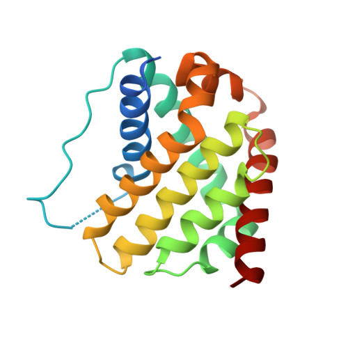
    Pro-apoptotic BAX is a cell fate regulator playing an important role in cellular homeostasis and pathological cell death. BAX is predominantly localized in the cytosol, where it has a quiescent monomer conformation. Following a pro-apoptotic trigger, cytosolic BAX is activated and translocates to the mitochondria to initiate mitochondrial dysfunction and apoptosis. Here, cellular, biochemical, and structural data unexpectedly demonstrate that cytosolic BAX also has an inactive dimer conformation that regulates its activation. The full-length crystal structure of the inactive BAX dimer revealed an asymmetric interaction consistent with inhibition of the N-terminal conformational change of one protomer and the displacement of the C-terminal helix α9 of the second protomer. This autoinhibited BAX dimer dissociates to BAX monomers before BAX can be activated. Our data support a model whereby the degree of apoptosis induction is regulated by the conformation of cytosolic BAX and identify an unprecedented mechanism of cytosolic BAX inhibition.


  • Organizational Affiliation

    Department of Biochemistry and Department of Medicine, Albert Einstein Cancer Center, Wilf Family Cardiovascular Research Institute, Albert Einstein College of Medicine, Bronx, NY 10461, USA.


Macromolecules
Find similar proteins by:  (by identity cutoff)  |  3D Structure
Entity ID: 1
MoleculeChains Sequence LengthOrganismDetailsImage
Apoptosis regulator BAX
A, B
192Homo sapiensMutation(s): 1 
Gene Names: BAXBCL2L4
UniProt & NIH Common Fund Data Resources
Find proteins for Q07812 (Homo sapiens)
Explore Q07812 
Go to UniProtKB:  Q07812
PHAROS:  Q07812
GTEx:  ENSG00000087088 
Entity Groups  
Sequence Clusters30% Identity50% Identity70% Identity90% Identity95% Identity100% Identity
UniProt GroupQ07812
Sequence Annotations
Expand
  • Reference Sequence
Experimental Data & Validation

Experimental Data

  • Method: X-RAY DIFFRACTION
  • Resolution: 3.25 Å
  • R-Value Free: 0.270 
  • R-Value Work: 0.211 
  • R-Value Observed: 0.214 
  • Space Group: P 1 21 1
Unit Cell:
Length ( Å )Angle ( ˚ )
a = 40.587α = 90
b = 65.01β = 89.87
c = 65.59γ = 90
Software Package:
Software NamePurpose
PHENIXrefinement
HKL-2000data reduction
HKL-2000data scaling
REFMACrefinement

Structure Validation

View Full Validation Report


Entry History 

Deposition Data

Revision History  (Full details and data files)

  • Version 1.0: 2016-07-20
    Type: Initial release
  • Version 1.1: 2016-08-17
    Changes: Database references, Derived calculations
  • Version 1.2: 2017-11-22
    Changes: Refinement description
  • Version 1.3: 2019-07-17
    Changes: Data collection, Refinement description
  • Version 1.4: 2023-09-20
    Changes: Data collection, Database references, Refinement description