4RZZ

Crystal structure of metallopeptidase-like dimethylsulphoniopropionate (DMSP) lyase RlDddP in complex with phosphate


Experimental Data Snapshot

  • Method: X-RAY DIFFRACTION
  • Resolution: 2.10 Å
  • R-Value Free: 0.194 
  • R-Value Work: 0.168 
  • R-Value Observed: 0.170 

wwPDB Validation   3D Report Full Report


This is version 1.2 of the entry. See complete history


Literature

Structural and molecular basis for the novel catalytic mechanism and evolution of DddP, an abundant peptidase-like bacterial Dimethylsulfoniopropionate lyase: a new enzyme from an old fold.

Wang, P.Chen, X.L.Li, C.Y.Gao, X.Zhu, D.Y.Xie, B.B.Qin, Q.L.Zhang, X.Y.Su, H.N.Zhou, B.C.Xun, L.Y.Zhang, Y.Z.

(2015) Mol Microbiol 98: 289-301

  • DOI: https://doi.org/10.1111/mmi.13119
  • Primary Citation of Related Structures:  
    4RZY, 4RZZ, 4S00, 4S01

  • PubMed Abstract: 

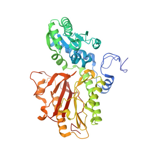
    The microbial cleavage of dimethylsulfoniopropionate (DMSP) generates volatile dimethyl sulfide (DMS) and is an important step in global sulfur and carbon cycles. DddP is a DMSP lyase in marine bacteria, and the deduced dddP gene product is abundant in marine metagenomic data sets. However, DddP belongs to the M24 peptidase family according to sequence alignment. Peptidases hydrolyze C-N bonds, but DddP is deduced to cleave C-S bonds. Mechanisms responsible for this striking functional shift are currently unknown. We determined the structures of DMSP lyase RlDddP (the DddP from Ruegeria lacuscaerulensis ITI_1157) bound to inhibitory 2-(N-morpholino) ethanesulfonic acid or PO4 (3-) and of two mutants of RlDddP bound to acrylate. Based on structural, mutational and biochemical analyses, we characterized a new ion-shift catalytic mechanism of RlDddP for DMSP cleavage. Furthermore, we suggested the structural mechanism leading to the loss of peptidase activity and the subsequent development of DMSP lyase activity in DddP. This study sheds light on the catalytic mechanism and the divergent evolution of DddP, leading to a better understanding of marine bacterial DMSP catabolism and global DMS production.


  • Organizational Affiliation

    State Key Laboratory of Microbial Technology, Shandong University, Jinan, 250100, China.


Macromolecules
Find similar proteins by:  (by identity cutoff)  |  3D Structure
Entity ID: 1
MoleculeChains Sequence LengthOrganismDetailsImage
Peptidase M24
A, B, C, D
447Ruegeria lacuscaerulensis ITI-1157Mutation(s): 0 
Gene Names: SL1157_2466
Entity Groups  
Sequence Clusters30% Identity50% Identity70% Identity90% Identity95% Identity100% Identity
Sequence Annotations
Expand
  • Reference Sequence
Small Molecules
Ligands 3 Unique
IDChains Name / Formula / InChI Key2D Diagram3D Interactions
PO4
Query on PO4

Download Ideal Coordinates CCD File 
G [auth A],
K [auth B],
P [auth C],
T [auth D]
PHOSPHATE ION
O4 P
NBIIXXVUZAFLBC-UHFFFAOYSA-K
GOL
Query on GOL

Download Ideal Coordinates CCD File 
H [auth A],
L [auth B],
M [auth B],
Q [auth C]
GLYCEROL
C3 H8 O3
PEDCQBHIVMGVHV-UHFFFAOYSA-N
FE
Query on FE

Download Ideal Coordinates CCD File 
E [auth A]
F [auth A]
I [auth B]
J [auth B]
N [auth C]
E [auth A],
F [auth A],
I [auth B],
J [auth B],
N [auth C],
O [auth C],
R [auth D],
S [auth D]
FE (III) ION
Fe
VTLYFUHAOXGGBS-UHFFFAOYSA-N
Experimental Data & Validation

Experimental Data

  • Method: X-RAY DIFFRACTION
  • Resolution: 2.10 Å
  • R-Value Free: 0.194 
  • R-Value Work: 0.168 
  • R-Value Observed: 0.170 
  • Space Group: P 32
Unit Cell:
Length ( Å )Angle ( ˚ )
a = 175.61α = 90
b = 175.61β = 90
c = 109.574γ = 120
Software Package:
Software NamePurpose
HKL-2000data collection
MLPHAREphasing
PHENIXrefinement
HKL-2000data reduction
HKL-2000data scaling

Structure Validation

View Full Validation Report


Entry History 

Deposition Data

Revision History  (Full details and data files)

  • Version 1.0: 2015-08-05
    Type: Initial release
  • Version 1.1: 2015-10-21
    Changes: Database references
  • Version 1.2: 2024-02-28
    Changes: Data collection, Database references, Derived calculations