4RU1

Crystal structure of carbohydrate transporter ACEI_1806 from Acidothermus cellulolyticus 11B, TARGET EFI-510965, in complex with myo-inositol


Experimental Data Snapshot

  • Method: X-RAY DIFFRACTION
  • Resolution: 1.50 Å
  • R-Value Free: 0.226 
  • R-Value Work: 0.185 
  • R-Value Observed: 0.187 

wwPDB Validation   3D Report Full Report


Ligand Structure Quality Assessment 


This is version 1.1 of the entry. See complete history


Literature

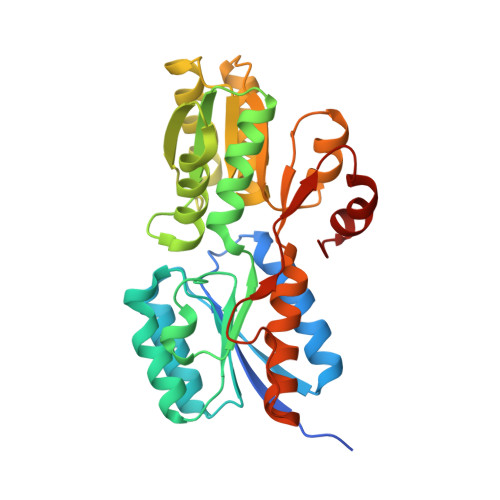
Crystal structure of carbohydrate transporter ACEI_1806 from Acidothermus cellulolyticus, TARGET EFI-510965.

Patskovsky, Y.Toro, R.Bhosle, R.Al Obaidi, N.Chamala, S.Scott Glenn, A.Attonito, J.D.Chowdhury, S.Lafleur, J.Siedel, R.D.Morisco, L.L.Wasserman, S.R.Hillerich, B.Love, J.Whalen, K.L.Gerlt, J.A.Almo, S.C.

To be published.

Macromolecules
Find similar proteins by:  (by identity cutoff)  |  3D Structure
Entity ID: 1
MoleculeChains Sequence LengthOrganismDetailsImage
Monosaccharide ABC transporter substrate-binding protein, CUT2 family
A, B, C, D, E
A, B, C, D, E, F, G, H, I, J, K, L
304Acidothermus cellulolyticus 11BMutation(s): 0 
Gene Names: Acel_1806
UniProt
Find proteins for A0LVW8 (Acidothermus cellulolyticus (strain ATCC 43068 / DSM 8971 / 11B))
Explore A0LVW8 
Go to UniProtKB:  A0LVW8
Entity Groups  
Sequence Clusters30% Identity50% Identity70% Identity90% Identity95% Identity100% Identity
UniProt GroupA0LVW8
Sequence Annotations
Expand
  • Reference Sequence
Small Molecules
Ligands 2 Unique
IDChains Name / Formula / InChI Key2D Diagram3D Interactions
CIT
Query on CIT

Download Ideal Coordinates CCD File 
O [auth C],
R [auth E]
CITRIC ACID
C6 H8 O7
KRKNYBCHXYNGOX-UHFFFAOYSA-N
INS
Query on INS

Download Ideal Coordinates CCD File 
M [auth A]
N [auth B]
P [auth C]
Q [auth D]
S [auth E]
M [auth A],
N [auth B],
P [auth C],
Q [auth D],
S [auth E],
T [auth F],
U [auth G],
V [auth H],
W [auth I],
X [auth J],
Y [auth K],
Z [auth L]
1,2,3,4,5,6-HEXAHYDROXY-CYCLOHEXANE
C6 H12 O6
CDAISMWEOUEBRE-GPIVLXJGSA-N
Experimental Data & Validation

Experimental Data

  • Method: X-RAY DIFFRACTION
  • Resolution: 1.50 Å
  • R-Value Free: 0.226 
  • R-Value Work: 0.185 
  • R-Value Observed: 0.187 
  • Space Group: P 1 21 1
Unit Cell:
Length ( Å )Angle ( ˚ )
a = 52.537α = 90
b = 149.976β = 91.85
c = 221.631γ = 90
Software Package:
Software NamePurpose
SHELXmodel building
ARP/wARPmodel building
REFMACrefinement
HKL-3000data reduction
HKL-3000data scaling
SHELXphasing

Structure Validation

View Full Validation Report


Ligand Structure Quality Assessment 


Entry History 

Revision History  (Full details and data files)

  • Version 1.0: 2014-12-17
    Type: Initial release
  • Version 1.1: 2018-01-24
    Changes: Structure summary