4RAP

Crystal structure of bacterial iron-containing dodecameric glycosyltransferase TibC from enterotoxigenic E.coli H10407


Experimental Data Snapshot

  • Method: X-RAY DIFFRACTION
  • Resolution: 2.88 Å
  • R-Value Free: 0.245 
  • R-Value Work: 0.208 
  • R-Value Observed: 0.210 

wwPDB Validation   3D Report Full Report


This is version 1.0 of the entry. See complete history


Literature

A structural mechanism for bacterial autotransporter glycosylation by a dodecameric heptosyltransferase family.

Yao, Q.Lu, Q.Wan, X.Song, F.Xu, Y.Hu, M.Zamyatina, A.Liu, X.Huang, N.Zhu, P.Shao, F.

(2014) Elife 3

  • DOI: https://doi.org/10.7554/eLife.03714
  • Primary Citation of Related Structures:  
    4RAP, 4RB4

  • PubMed Abstract: 

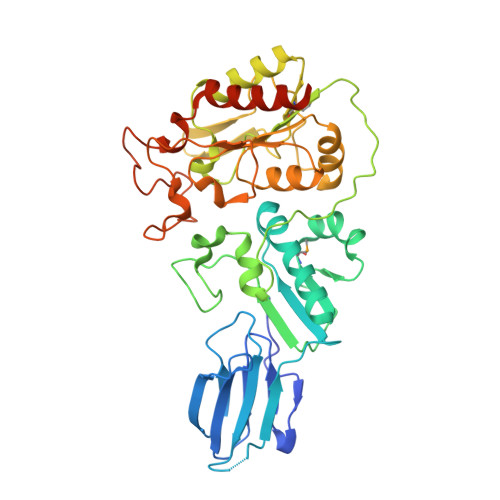
    A large group of bacterial virulence autotransporters including AIDA-I from diffusely adhering E. coli (DAEC) and TibA from enterotoxigenic E. coli (ETEC) require hyperglycosylation for functioning. Here we demonstrate that TibC from ETEC harbors a heptosyltransferase activity on TibA and AIDA-I, defining a large family of bacterial autotransporter heptosyltransferases (BAHTs). The crystal structure of TibC reveals a characteristic ring-shape dodecamer. The protomer features an N-terminal β-barrel, a catalytic domain, a β-hairpin thumb, and a unique iron-finger motif. The iron-finger motif contributes to back-to-back dimerization; six dimers form the ring through β-hairpin thumb-mediated hand-in-hand contact. The structure of ADP-D-glycero-β-D-manno-heptose (ADP-D,D-heptose)-bound TibC reveals a sugar transfer mechanism and also the ligand stereoselectivity determinant. Electron-cryomicroscopy analyses uncover a TibC-TibA dodecamer/hexamer assembly with two enzyme molecules binding to one TibA substrate. The complex structure also highlights a high efficient hyperglycosylation of six autotransporter substrates simultaneously by the dodecamer enzyme complex.


  • Organizational Affiliation

    Dr Feng Shao's Laboratory, National Institute of Biological Sciences, Beijing, China.


Macromolecules
Find similar proteins by:  (by identity cutoff)  |  3D Structure
Entity ID: 1
MoleculeChains Sequence LengthOrganismDetailsImage
Glycosyltransferase TibC
A, B, C, D, E
A, B, C, D, E, F, G, H, I, J, K, L
406Escherichia coli ETEC H10407Mutation(s): 6 
Gene Names: tibCETEC_2140
EC: 2.4
UniProt
Find proteins for Q9S4K6 (Escherichia coli O78:H11 (strain H10407 / ETEC))
Explore Q9S4K6 
Go to UniProtKB:  Q9S4K6
Entity Groups  
Sequence Clusters30% Identity50% Identity70% Identity90% Identity95% Identity100% Identity
UniProt GroupQ9S4K6
Sequence Annotations
Expand
  • Reference Sequence
Small Molecules
Ligands 2 Unique
IDChains Name / Formula / InChI Key2D Diagram3D Interactions
EDO
Query on EDO

Download Ideal Coordinates CCD File 
CA [auth L],
P [auth C],
W [auth I],
X [auth I],
Z [auth J]
1,2-ETHANEDIOL
C2 H6 O2
LYCAIKOWRPUZTN-UHFFFAOYSA-N
FE
Query on FE

Download Ideal Coordinates CCD File 
AA [auth K]
BA [auth L]
M [auth A]
N [auth B]
O [auth C]
AA [auth K],
BA [auth L],
M [auth A],
N [auth B],
O [auth C],
Q [auth D],
R [auth E],
S [auth F],
T [auth G],
U [auth H],
V [auth I],
Y [auth J]
FE (III) ION
Fe
VTLYFUHAOXGGBS-UHFFFAOYSA-N
Modified Residues  1 Unique
IDChains TypeFormula2D DiagramParent
MSE
Query on MSE
A, B, C, D, E
A, B, C, D, E, F, G, H, I, J, K, L
L-PEPTIDE LINKINGC5 H11 N O2 SeMET
Experimental Data & Validation

Experimental Data

  • Method: X-RAY DIFFRACTION
  • Resolution: 2.88 Å
  • R-Value Free: 0.245 
  • R-Value Work: 0.208 
  • R-Value Observed: 0.210 
  • Space Group: P 1 21 1
Unit Cell:
Length ( Å )Angle ( ˚ )
a = 87.794α = 90
b = 314.368β = 101.44
c = 164.527γ = 90
Software Package:
Software NamePurpose
Blu-Icedata collection
SHELXSphasing
PHENIXrefinement
HKL-2000data reduction
HKL-2000data scaling

Structure Validation

View Full Validation Report


Entry History 

Deposition Data

Revision History  (Full details and data files)

  • Version 1.0: 2014-10-29
    Type: Initial release