4RAF

Crystal structure of PP2Ca-D38A


Experimental Data Snapshot

  • Method: X-RAY DIFFRACTION
  • Resolution: 2.00 Å
  • R-Value Free: 0.222 
  • R-Value Work: 0.194 
  • R-Value Observed: 0.196 

wwPDB Validation   3D Report Full Report


This is version 1.1 of the entry. See complete history


Literature

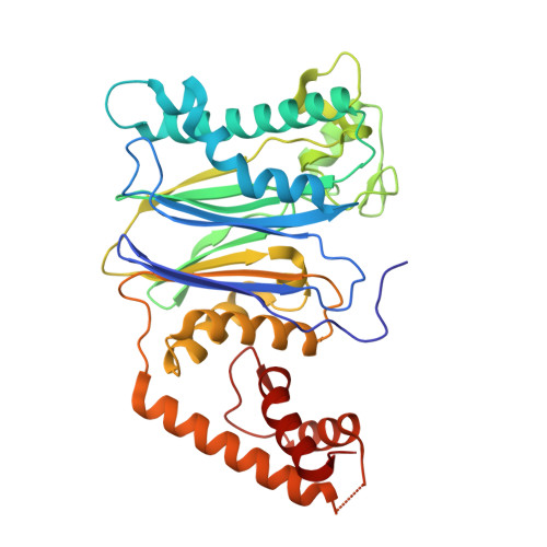
The catalytic role of the M2 metal ion in PP2C alpha

Pan, C.Tang, J.Y.Xu, Y.F.Xiao, P.Liu, H.D.Wang, H.A.Wang, W.B.Meng, F.G.Yu, X.Sun, J.P.

(2015) Sci Rep 5: 8560-8560

  • DOI: https://doi.org/10.1038/srep08560
  • Primary Citation of Related Structures:  
    4RAF, 4RAG

  • PubMed Abstract: 

    PP2C family phosphatases (the type 2C family of protein phosphatases; or metal-dependent phosphatase, PPM) constitute an important class of signaling enzymes that regulate many fundamental life activities. All PP2C family members have a conserved binuclear metal ion active center that is essential for their catalysis. However, the catalytic role of each metal ion during catalysis remains elusive. In this study, we discovered that mutations in the structurally buried D38 residue of PP2Cα (PPM1A) redefined the water-mediated hydrogen network in the active site and selectively disrupted M2 metal ion binding. Using the D38A and D38K mutations of PP2Cα as specific tools in combination with enzymology analysis, our results demonstrated that the M2 metal ion determines the rate-limiting step of substrate hydrolysis, participates in dianion substrate binding and stabilizes the leaving group after P-O bond cleavage. The newly characterized catalytic role of the M2 metal ion in this family not only provides insight into how the binuclear metal centers of the PP2C phosphatases are organized for efficient catalysis but also helps increase our understanding of the function and substrate specificity of PP2C family members.


  • Organizational Affiliation

    1] Key Laboratory Experimental Teratology of the Ministry of Education and Department of Biochemistry and Molecular Biology, Shandong University, School of Medicine, Jinan, Shandong, China [2] Qilu Hospital of Shandong University, Jinan, China.


Macromolecules
Find similar proteins by:  (by identity cutoff)  |  3D Structure
Entity ID: 1
MoleculeChains Sequence LengthOrganismDetailsImage
Protein phosphatase 1A367Homo sapiensMutation(s): 1 
Gene Names: PPM1APPPM1A
EC: 3.1.3.16
UniProt & NIH Common Fund Data Resources
Find proteins for P35813 (Homo sapiens)
Explore P35813 
Go to UniProtKB:  P35813
PHAROS:  P35813
GTEx:  ENSG00000100614 
Entity Groups  
Sequence Clusters30% Identity50% Identity70% Identity90% Identity95% Identity100% Identity
UniProt GroupP35813
Sequence Annotations
Expand
  • Reference Sequence
Small Molecules
Ligands 1 Unique
IDChains Name / Formula / InChI Key2D Diagram3D Interactions
MN
Query on MN

Download Ideal Coordinates CCD File 
B [auth A]MANGANESE (II) ION
Mn
WAEMQWOKJMHJLA-UHFFFAOYSA-N
Experimental Data & Validation

Experimental Data

  • Method: X-RAY DIFFRACTION
  • Resolution: 2.00 Å
  • R-Value Free: 0.222 
  • R-Value Work: 0.194 
  • R-Value Observed: 0.196 
  • Space Group: P 31 2 1
Unit Cell:
Length ( Å )Angle ( ˚ )
a = 91.262α = 90
b = 91.262β = 90
c = 105.767γ = 120
Software Package:
Software NamePurpose
HKL-2000data collection
PHASESphasing
PHENIXrefinement
HKL-2000data reduction
HKL-2000data scaling

Structure Validation

View Full Validation Report


Entry History 

Deposition Data

Revision History  (Full details and data files)

  • Version 1.0: 2015-08-26
    Type: Initial release
  • Version 1.1: 2023-11-08
    Changes: Data collection, Database references, Derived calculations, Refinement description