4QZV

Bat-derived coronavirus HKU4 uses MERS-CoV receptor human CD26 for cell entry


Experimental Data Snapshot

  • Method: X-RAY DIFFRACTION
  • Resolution: 2.59 Å
  • R-Value Free: 0.224 
  • R-Value Work: 0.190 
  • R-Value Observed: 0.191 

wwPDB Validation   3D Report Full Report


Ligand Structure Quality Assessment 


This is version 2.0 of the entry. See complete history


Literature

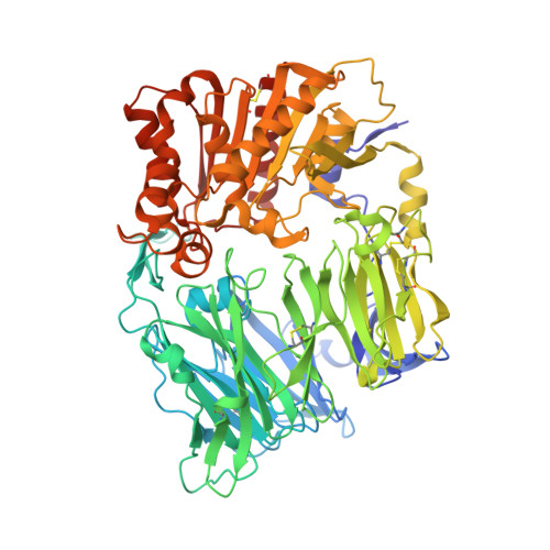
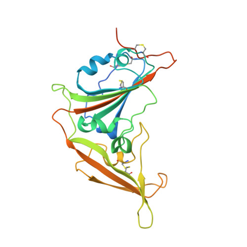
Bat Origins of MERS-CoV Supported by Bat Coronavirus HKU4 Usage of Human Receptor CD26.

Wang, Q.H.Qi, J.X.Yuan, Y.Xuan, Y.Han, P.Wan, Y.Ji, W.Li, Y.Wu, Y.Wang, J.Iwamoto, A.Woo, P.C.Yuen, K.Y.Yan, J.Lu, G.W.Gao, G.F.

(2014) Cell Host Microbe 16: 328-337

  • DOI: https://doi.org/10.1016/j.chom.2014.08.009
  • Primary Citation of Related Structures:  
    4QZV

  • PubMed Abstract: 

    The recently reported Middle East respiratory syndrome coronavirus (MERS-CoV) is phylogenetically closely related to the bat coronaviruses (BatCoVs) HKU4 and HKU5. However, the evolutionary pathway of MERS-CoV is still unclear. A receptor binding domain (RBD) in the MERS-CoV envelope-embedded spike protein specifically engages human CD26 (hCD26) to initiate viral entry. The high sequence identity in the viral spike protein prompted us to investigate if HKU4 and HKU5 can recognize hCD26 for cell entry. We found that HKU4-RBD, but not HKU5-RBD, binds to hCD26, and pseudotyped viruses embedding HKU4 spike can infect cells via hCD26 recognition. The structure of the HKU4-RBD/hCD26 complex revealed a hCD26-binding mode similar overall to that observed for MERS-RBD. HKU4-RBD, however, is less adapted to hCD26 than MERS-RBD, explaining its lower affinity for receptor binding. Our findings support a bat origin for MERS-CoV and indicate the need for surveillance of HKU4-related viruses in bats.


  • Organizational Affiliation

    CAS Key Laboratory of Pathogenic Microbiology and Immunology, Institute of Microbiology, Chinese Academy of Sciences, Beijing 100101, China.


Macromolecules
Find similar proteins by:  (by identity cutoff)  |  3D Structure
Entity ID: 1
MoleculeChains Sequence LengthOrganismDetailsImage
Dipeptidyl peptidase 4
A, C
734Homo sapiensMutation(s): 0 
Gene Names: DPP4ADCP2CD26
EC: 3.4.14.5
UniProt & NIH Common Fund Data Resources
Find proteins for P27487 (Homo sapiens)
Explore P27487 
Go to UniProtKB:  P27487
PHAROS:  P27487
GTEx:  ENSG00000197635 
Entity Groups  
Sequence Clusters30% Identity50% Identity70% Identity90% Identity95% Identity100% Identity
UniProt GroupP27487
Sequence Annotations
Expand
  • Reference Sequence
Find similar proteins by:  (by identity cutoff)  |  3D Structure
Entity ID: 2
MoleculeChains Sequence LengthOrganismDetailsImage
Spike protein S1
B, D
246Tylonycteris bat coronavirus HKU4Mutation(s): 0 
Gene Names: S2
UniProt
Find proteins for A3EX94 (Bat coronavirus HKU4)
Explore A3EX94 
Go to UniProtKB:  A3EX94
Entity Groups  
Sequence Clusters30% Identity50% Identity70% Identity90% Identity95% Identity100% Identity
UniProt GroupA3EX94
Sequence Annotations
Expand
  • Reference Sequence
Oligosaccharides

Help

Entity ID: 3
MoleculeChains Length2D Diagram Glycosylation3D Interactions
2-acetamido-2-deoxy-beta-D-glucopyranose-(1-4)-2-acetamido-2-deoxy-beta-D-glucopyranose
E, G, H, I, K
E, G, H, I, K, L
2N-Glycosylation
Glycosylation Resources
GlyTouCan:  G42666HT
GlyCosmos:  G42666HT
GlyGen:  G42666HT
Entity ID: 4
MoleculeChains Length2D Diagram Glycosylation3D Interactions
beta-D-mannopyranose-(1-4)-2-acetamido-2-deoxy-beta-D-glucopyranose-(1-4)-2-acetamido-2-deoxy-beta-D-glucopyranose
F, J
3N-Glycosylation
Glycosylation Resources
GlyTouCan:  G15407YE
GlyCosmos:  G15407YE
GlyGen:  G15407YE
Experimental Data & Validation

Experimental Data

  • Method: X-RAY DIFFRACTION
  • Resolution: 2.59 Å
  • R-Value Free: 0.224 
  • R-Value Work: 0.190 
  • R-Value Observed: 0.191 
  • Space Group: P 2 21 21
Unit Cell:
Length ( Å )Angle ( ˚ )
a = 78.801α = 90
b = 203.166β = 90
c = 207.753γ = 90
Software Package:
Software NamePurpose
Blu-Icedata collection
PHASERphasing
PHENIXrefinement
HKL-2000data reduction
HKL-2000data scaling

Structure Validation

View Full Validation Report


Ligand Structure Quality Assessment 


Entry History 

Deposition Data

Revision History  (Full details and data files)

  • Version 1.0: 2014-10-29
    Type: Initial release
  • Version 2.0: 2020-07-29
    Type: Remediation
    Reason: Carbohydrate remediation
    Changes: Advisory, Atomic model, Data collection, Database references, Derived calculations, Structure summary