4QUM

Crystal structure of PTPN3 (PTPH1) in complex with a dually phosphorylated MAPK12 peptide


Experimental Data Snapshot

  • Method: X-RAY DIFFRACTION
  • Resolution: 2.52 Å
  • R-Value Free: 0.245 
  • R-Value Work: 0.197 
  • R-Value Observed: 0.199 

wwPDB Validation   3D Report Full Report


This is version 1.3 of the entry. See complete history


Literature

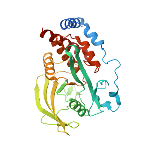
Reciprocal allosteric regulation of p38 gamma and PTPN3 involves a PDZ domain-modulated complex formation.

Chen, K.E.Lin, S.Y.Wu, M.J.Ho, M.R.Santhanam, A.Chou, C.C.Meng, T.C.Wang, A.H.J.

(2014) Sci Signal 7: ra98-ra98

  • DOI: https://doi.org/10.1126/scisignal.2005722
  • Primary Citation of Related Structures:  
    4QUM, 4QUN

  • PubMed Abstract: 

    The mitogen-activated protein kinase p38γ (also known as MAPK12) and its specific phosphatase PTPN3 (also known as PTPH1) cooperate to promote Ras-induced oncogenesis. We determined the architecture of the PTPN3-p38γ complex by a hybrid method combining x-ray crystallography, small-angle x-ray scattering, and chemical cross-linking coupled to mass spectrometry. A unique feature of the glutamic acid-containing loop (E-loop) of the phosphatase domain defined the substrate specificity of PTPN3 toward fully activated p38γ. The solution structure revealed the formation of an active-state complex between p38γ and the phosphatase domain of PTPN3. The PDZ domain of PTPN3 stabilized the active-state complex through an interaction with the PDZ-binding motif of p38γ. This interaction alleviated autoinhibition of PTPN3, enabling efficient tyrosine dephosphorylation of p38γ. Our findings may enable structure-based drug design targeting the PTPN3-p38γ interaction as an anticancer therapeutic.


  • Organizational Affiliation

    Institute of Biological Chemistry, Academia Sinica, Taipei 11581, Taiwan.


Macromolecules
Find similar proteins by:  (by identity cutoff)  |  3D Structure
Entity ID: 1
MoleculeChains Sequence LengthOrganismDetailsImage
Tyrosine-protein phosphatase non-receptor type 3306Homo sapiensMutation(s): 2 
Gene Names: PTPH1PTPN3
EC: 3.1.3.48
UniProt & NIH Common Fund Data Resources
Find proteins for P26045 (Homo sapiens)
Explore P26045 
Go to UniProtKB:  P26045
PHAROS:  P26045
GTEx:  ENSG00000070159 
Entity Groups  
Sequence Clusters30% Identity50% Identity70% Identity90% Identity95% Identity100% Identity
UniProt GroupP26045
Sequence Annotations
Expand
  • Reference Sequence

Find similar proteins by:  Sequence   |   3D Structure  

Entity ID: 2
MoleculeChains Sequence LengthOrganismDetailsImage
Mitogen-activated protein kinase 129Homo sapiensMutation(s): 1 
EC: 2.7.11.24
UniProt & NIH Common Fund Data Resources
Find proteins for P53778 (Homo sapiens)
Explore P53778 
Go to UniProtKB:  P53778
PHAROS:  P53778
GTEx:  ENSG00000188130 
Entity Groups  
UniProt GroupP53778
Sequence Annotations
Expand
  • Reference Sequence
Small Molecules
Modified Residues  2 Unique
IDChains TypeFormula2D DiagramParent
PTR
Query on PTR
B
L-PEPTIDE LINKINGC9 H12 N O6 PTYR
TPO
Query on TPO
B
L-PEPTIDE LINKINGC4 H10 N O6 PTHR
Experimental Data & Validation

Experimental Data

  • Method: X-RAY DIFFRACTION
  • Resolution: 2.52 Å
  • R-Value Free: 0.245 
  • R-Value Work: 0.197 
  • R-Value Observed: 0.199 
  • Space Group: P 31 2 1
Unit Cell:
Length ( Å )Angle ( ˚ )
a = 75.491α = 90
b = 75.491β = 90
c = 109.605γ = 120
Software Package:
Software NamePurpose
Blu-Icedata collection
PHASESphasing
PHENIXrefinement
HKL-2000data reduction
HKL-2000data scaling

Structure Validation

View Full Validation Report


Entry History 

Deposition Data

Revision History  (Full details and data files)

  • Version 1.0: 2014-12-10
    Type: Initial release
  • Version 1.1: 2018-07-18
    Changes: Advisory, Data collection
  • Version 1.2: 2023-11-08
    Changes: Advisory, Data collection, Database references, Derived calculations, Refinement description
  • Version 1.3: 2023-12-06
    Changes: Data collection