Structural basis of substrate selectivity of E. coli prolidase.
Weaver, J., Watts, T., Li, P., Rye, H.S.(2014) PLoS One 9: e111531-e111531
- PubMed: 25354344
- DOI: https://doi.org/10.1371/journal.pone.0111531
- Primary Citation of Related Structures:
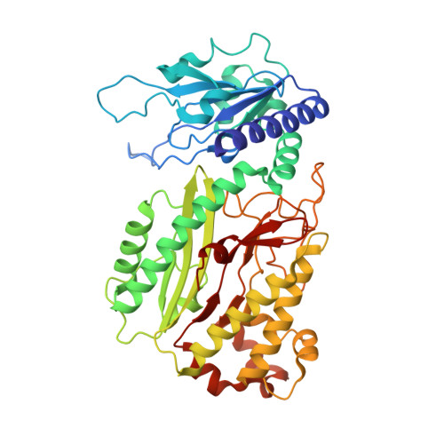
4QR8 - PubMed Abstract:
Prolidases, metalloproteases that catalyze the cleavage of Xaa-Pro dipeptides, are conserved enzymes found in prokaryotes and eukaryotes. In humans, prolidase is crucial for the recycling of collagen. To further characterize the essential elements of this enzyme, we utilized the Escherichia coli prolidase, PepQ, which shares striking similarity with eukaryotic prolidases. Through structural and bioinformatic insights, we have extended previous characterizations of the prolidase active site, uncovering a key component for substrate specificity. Here we report the structure of E. coli PepQ, solved at 2.0 Å resolution. The structure shows an antiparallel, dimeric protein, with each subunit containing N-terminal and C-terminal domains. The C-terminal domain is formed by the pita-bread fold typical for this family of metalloproteases, with two Mg(II) ions coordinated by five amino-acid ligands. Comparison of the E. coli PepQ structure and sequence with homologous structures and sequences from a diversity of organisms reveals distinctions between prolidases from Gram-positive eubacteria and archaea, and those from Gram-negative eubacteria, including the presence of loop regions in the E. coli protein that are conserved in eukaryotes. One such loop contains a completely conserved arginine near the catalytic site. This conserved arginine is predicted by docking simulations to interact with the C-terminus of the substrate dipeptide. Kinetic analysis using both a charge-neutralized substrate and a charge-reversed variant of PepQ support this conclusion, and allow for the designation of a new role for this key region of the enzyme active site.
Organizational Affiliation:
Department of Biochemistry and Biophysics, Texas A&M University, College Station, Texas, United States of America.