4QHI

Crystal structure of Methanocaldococcus jannaschii selecase mutant R36W


Experimental Data Snapshot

  • Method: X-RAY DIFFRACTION
  • Resolution: 2.30 Å
  • R-Value Free: 0.247 
  • R-Value Work: 0.222 
  • R-Value Observed: 0.222 

wwPDB Validation   3D Report Full Report


This is version 1.3 of the entry. See complete history


Literature

Multiple stable conformations account for reversible concentration-dependent oligomerization and autoinhibition of a metamorphic metallopeptidase

Lopez-Pelegrin, M.Cerda-Costa, N.Cintas-Pedrola, A.Herranz-Trillo, F.Bernado, P.Peinado, J.R.Arolas, J.L.Gomis-Ruth, F.X.

(2014) Angew Chem Int Ed Engl 53: 10624-10630

  • DOI: https://doi.org/10.1002/anie.201405727
  • Primary Citation of Related Structures:  
    4QHF, 4QHG, 4QHH, 4QHI, 4QHJ

  • PubMed Abstract: 

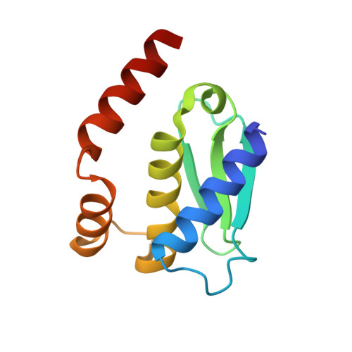
    Molecular plasticity controls enzymatic activity: the native fold of a protein in a given environment is normally unique and at a global free-energy minimum. Some proteins, however, spontaneously undergo substantial fold switching to reversibly transit between defined conformers, the "metamorphic" proteins. Here, we present a minimal metamorphic, selective, and specific caseinolytic metallopeptidase, selecase, which reversibly transits between several different states of defined three-dimensional structure, which are associated with loss of enzymatic activity due to autoinhibition. The latter is triggered by sequestering the competent conformation in incompetent but structured dimers, tetramers, and octamers. This system, which is compatible with a discrete multifunnel energy landscape, affords a switch that provides a reversible mechanism of control of catalytic activity unique in nature.


  • Organizational Affiliation

    Proteolysis Lab, Molecular Biology Institute of Barcelona, CSIC, Barcelona Science Park c/Baldiri Reixac, 15-21, 08028 Barcelona (Spain) http://www.ibmb.csic.es/home/xgomis.


Macromolecules
Find similar proteins by:  (by identity cutoff)  |  3D Structure
Entity ID: 1
MoleculeChains Sequence LengthOrganismDetailsImage
Uncharacterized protein MJ1213
A, B, C, D
110Methanocaldococcus jannaschii DSM 2661Mutation(s): 1 
Gene Names: MJ1213
UniProt
Find proteins for Q58610 (Methanocaldococcus jannaschii (strain ATCC 43067 / DSM 2661 / JAL-1 / JCM 10045 / NBRC 100440))
Explore Q58610 
Go to UniProtKB:  Q58610
Entity Groups  
Sequence Clusters30% Identity50% Identity70% Identity90% Identity95% Identity100% Identity
UniProt GroupQ58610
Sequence Annotations
Expand
  • Reference Sequence
Small Molecules
Ligands 3 Unique
IDChains Name / Formula / InChI Key2D Diagram3D Interactions
GOL
Query on GOL

Download Ideal Coordinates CCD File 
F [auth A],
I [auth B],
J [auth B],
L [auth C]
GLYCEROL
C3 H8 O3
PEDCQBHIVMGVHV-UHFFFAOYSA-N
ZN
Query on ZN

Download Ideal Coordinates CCD File 
E [auth A],
H [auth B],
K [auth C],
M [auth D]
ZINC ION
Zn
PTFCDOFLOPIGGS-UHFFFAOYSA-N
CL
Query on CL

Download Ideal Coordinates CCD File 
G [auth A]CHLORIDE ION
Cl
VEXZGXHMUGYJMC-UHFFFAOYSA-M
Experimental Data & Validation

Experimental Data

  • Method: X-RAY DIFFRACTION
  • Resolution: 2.30 Å
  • R-Value Free: 0.247 
  • R-Value Work: 0.222 
  • R-Value Observed: 0.222 
  • Space Group: P 1 21 1
Unit Cell:
Length ( Å )Angle ( ˚ )
a = 37.69α = 90
b = 101.09β = 99.83
c = 76.47γ = 90
Software Package:
Software NamePurpose
MOLREPphasing
BUSTERrefinement
XDSdata reduction
XSCALEdata scaling

Structure Validation

View Full Validation Report


Entry History 

Revision History  (Full details and data files)

  • Version 1.0: 2014-07-16
    Type: Initial release
  • Version 1.1: 2014-10-22
    Changes: Database references
  • Version 1.2: 2024-03-20
    Changes: Data collection, Database references, Derived calculations
  • Version 1.3: 2024-04-03
    Changes: Refinement description