


Crystal structure of phosphoglucomutase from Leishmania major at 3.5 angstrom resolution
Waugh, B., Sen, U., Banerjee, R.To be published.
Experimental Data Snapshot
wwPDB Validation 3D Report Full Report

Entity ID: 1 | |||||
|---|---|---|---|---|---|
| Molecule | Chains | Sequence Length | Organism | Details | Image |
| Putative phosphoglucomutase | 592 | Leishmania major | Mutation(s): 0 Gene Names: LMJF_21_0640 EC: 5.4.2.2 | 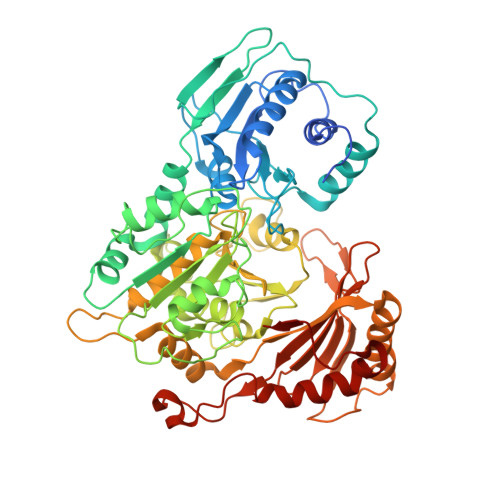 | |
UniProt | |||||
Find proteins for Q4QCF1 (Leishmania major) Explore Q4QCF1 Go to UniProtKB: Q4QCF1 | |||||
Entity Groups | |||||
| Sequence Clusters | 30% Identity50% Identity70% Identity90% Identity95% Identity100% Identity | ||||
| UniProt Group | Q4QCF1 | ||||
Sequence AnnotationsExpand | |||||
| |||||
| Ligands 1 Unique | |||||
|---|---|---|---|---|---|
| ID | Chains | Name / Formula / InChI Key | 2D Diagram | 3D Interactions | |
| MG Query on MG | E [auth A], F [auth B], G [auth C], H [auth D] | MAGNESIUM ION Mg JLVVSXFLKOJNIY-UHFFFAOYSA-N |  | ||
| Length ( Å ) | Angle ( ˚ ) |
|---|---|
| a = 202.358 | α = 90 |
| b = 114.971 | β = 110.03 |
| c = 125.915 | γ = 90 |
| Software Name | Purpose |
|---|---|
| Aimless | data scaling |
| PHASER | phasing |
| PHENIX | refinement |
| PDB_EXTRACT | data extraction |
| XDS | data reduction |
RCSB PDB (citation) is hosted by
RCSB PDB is a member of the

