4Q35

Structure of a membrane protein


Experimental Data Snapshot

  • Method: X-RAY DIFFRACTION
  • Resolution: 2.39 Å
  • R-Value Free: 0.231 
  • R-Value Work: 0.186 
  • R-Value Observed: 0.189 

wwPDB Validation   3D Report Full Report


Ligand Structure Quality Assessment 


This is version 1.2 of the entry. See complete history


Literature

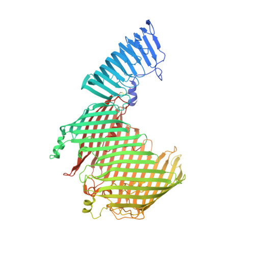
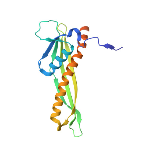
Structural basis for lipopolysaccharide insertion in the bacterial outer membrane.

Qiao, S.Luo, Q.Zhao, Y.Zhang, X.C.Huang, Y.

(2014) Nature 511: 108-111

  • DOI: https://doi.org/10.1038/nature13484
  • Primary Citation of Related Structures:  
    4Q35

  • PubMed Abstract: 

    One of the fundamental properties of biological membranes is the asymmetric distribution of membrane lipids. In Gram-negative bacteria, the outer leaflet of the outer membrane is composed predominantly of lipopolysaccharides (LPS). The export of LPS requires seven essential lipopolysaccharide transport (Lpt) proteins to move LPS from the inner membrane, through the periplasm to the surface. Of the seven Lpt proteins, the LptD-LptE complex is responsible for inserting LPS into the external leaflet of the outer membrane. Here we report the crystal structure of the ∼110-kilodalton membrane protein complex LptD-LptE from Shigella flexneri at 2.4 Å resolution. The structure reveals an unprecedented two-protein plug-and-barrel architecture with LptE embedded into a 26-stranded β-barrel formed by LptD. Importantly, the secondary structures of the first two β-strands are distorted by two proline residues, weakening their interactions with neighbouring β-strands and creating a potential portal on the barrel wall that could allow lateral diffusion of LPS into the outer membrane. The crystal structure of the LptD-LptE complex opens the door to new antibiotic strategies targeting the bacterial outer membrane.


  • Organizational Affiliation

    1] National Laboratory of Biomacromolecules, National Center of Protein Science-Beijing, Institute of Biophysics, Chinese Academy of Sciences, Beijing 100101, China [2] University of Chinese Academy of Sciences, Beijing 100101, China.


Macromolecules
Find similar proteins by:  (by identity cutoff)  |  3D Structure
Entity ID: 1
MoleculeChains Sequence LengthOrganismDetailsImage
LPS-assembly protein LptD802Shigella flexneriMutation(s): 0 
Gene Names: lptDimpostASF0051S0053
Membrane Entity: Yes 
UniProt
Find proteins for Q83SQ0 (Shigella flexneri)
Explore Q83SQ0 
Go to UniProtKB:  Q83SQ0
Entity Groups  
Sequence Clusters30% Identity50% Identity70% Identity90% Identity95% Identity100% Identity
UniProt GroupQ83SQ0
Sequence Annotations
Expand
  • Reference Sequence
Find similar proteins by:  (by identity cutoff)  |  3D Structure
Entity ID: 2
MoleculeChains Sequence LengthOrganismDetailsImage
LPS-assembly lipoprotein LptE175Shigella flexneriMutation(s): 0 
Gene Names: lptErlpBSF0640S0662
Membrane Entity: Yes 
UniProt
Find proteins for Q83LX4 (Shigella flexneri)
Explore Q83LX4 
Go to UniProtKB:  Q83LX4
Entity Groups  
Sequence Clusters30% Identity50% Identity70% Identity90% Identity95% Identity100% Identity
UniProt GroupQ83LX4
Sequence Annotations
Expand
  • Reference Sequence
Small Molecules
Ligands 3 Unique
IDChains Name / Formula / InChI Key2D Diagram3D Interactions
C8E
Query on C8E

Download Ideal Coordinates CCD File 
C [auth A]
D [auth A]
E [auth A]
I [auth A]
K [auth A]
C [auth A],
D [auth A],
E [auth A],
I [auth A],
K [auth A],
M [auth A]
(HYDROXYETHYLOXY)TRI(ETHYLOXY)OCTANE
C16 H34 O5
FEOZZFHAVXYAMB-UHFFFAOYSA-N
LDA
Query on LDA

Download Ideal Coordinates CCD File 
F [auth A]
G [auth A]
H [auth A]
J [auth A]
L [auth A]
F [auth A],
G [auth A],
H [auth A],
J [auth A],
L [auth A],
N [auth A],
O [auth A]
LAURYL DIMETHYLAMINE-N-OXIDE
C14 H31 N O
SYELZBGXAIXKHU-UHFFFAOYSA-N
SO4
Query on SO4

Download Ideal Coordinates CCD File 
P [auth A]
Q [auth A]
R [auth A]
S [auth A]
T [auth A]
P [auth A],
Q [auth A],
R [auth A],
S [auth A],
T [auth A],
U [auth A],
V [auth A],
W [auth A],
X [auth B]
SULFATE ION
O4 S
QAOWNCQODCNURD-UHFFFAOYSA-L
Experimental Data & Validation

Experimental Data

  • Method: X-RAY DIFFRACTION
  • Resolution: 2.39 Å
  • R-Value Free: 0.231 
  • R-Value Work: 0.186 
  • R-Value Observed: 0.189 
  • Space Group: C 1 2 1
Unit Cell:
Length ( Å )Angle ( ˚ )
a = 240.823α = 90
b = 116.62β = 103.71
c = 68.038γ = 90
Software Package:
Software NamePurpose
PHASESphasing
PHENIXrefinement
DENZOdata reduction
SCALEPACKdata scaling

Structure Validation

View Full Validation Report


Ligand Structure Quality Assessment 


Entry History 

Deposition Data

Revision History  (Full details and data files)

  • Version 1.0: 2014-06-25
    Type: Initial release
  • Version 1.1: 2014-07-09
    Changes: Database references
  • Version 1.2: 2019-12-25
    Changes: Database references