4PQQ

The crystal structure of discoidin domain from muskelin


Experimental Data Snapshot

  • Method: X-RAY DIFFRACTION
  • Resolution: 1.55 Å
  • R-Value Free: 0.176 
  • R-Value Work: 0.153 
  • R-Value Observed: 0.155 

wwPDB Validation   3D Report Full Report


This is version 1.2 of the entry. See complete history


Literature

Structure of mouse muskelin discoidin domain and biochemical characterization of its self-association.

Kim, K.H.Hong, S.K.Hwang, K.Y.Kim, E.E.

(2014) Acta Crystallogr D Biol Crystallogr 70: 2863-2874

  • DOI: https://doi.org/10.1107/S139900471401894X
  • Primary Citation of Related Structures:  
    4PQQ

  • PubMed Abstract: 

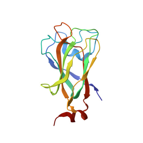
    Muskelin is an intracellular kelch-repeat protein comprised of discoidin, LisH, CTLH and kelch-repeat domains. It is involved in cell adhesion and the regulation of cytoskeleton dynamics as well as being a component of a putative E3 ligase complex. Here, the first crystal structure of mouse muskelin discoidin domain (MK-DD) is reported at 1.55 Å resolution, which reveals a distorted eight-stranded β-barrel with two short α-helices at one end of the barrel. Interestingly, the N- and C-termini are not linked by the disulfide bonds found in other eukaryotic discoidin structures. A highly conserved MIND motif appears to be the determinant for MK-DD specific interaction together with the spike loops. Analysis of interdomain interaction shows that MK-DD binds the kelch-repeat domain directly and that this interaction depends on the presence of the LisH domain.


  • Organizational Affiliation

    Biomedical Research Institute, Korea Institute of Science and Technology, Hwarang-ro 14-gil 5, Seongbuk-gu, Seoul 136-791, Republic of Korea.


Macromolecules
Find similar proteins by:  (by identity cutoff)  |  3D Structure
Entity ID: 1
MoleculeChains Sequence LengthOrganismDetailsImage
Muskelin156Mus musculusMutation(s): 0 
Gene Names: Mkln1
UniProt
Find proteins for O89050 (Mus musculus)
Explore O89050 
Go to UniProtKB:  O89050
Entity Groups  
Sequence Clusters30% Identity50% Identity70% Identity90% Identity95% Identity100% Identity
UniProt GroupO89050
Sequence Annotations
Expand
  • Reference Sequence
Experimental Data & Validation

Experimental Data

  • Method: X-RAY DIFFRACTION
  • Resolution: 1.55 Å
  • R-Value Free: 0.176 
  • R-Value Work: 0.153 
  • R-Value Observed: 0.155 
  • Space Group: H 3
Unit Cell:
Length ( Å )Angle ( ˚ )
a = 98.303α = 90
b = 98.303β = 90
c = 57.317γ = 120
Software Package:
Software NamePurpose
ADSCdata collection
SOLVEphasing
PHENIXrefinement
HKL-2000data reduction
HKL-2000data scaling

Structure Validation

View Full Validation Report


Entry History 

Deposition Data

Revision History  (Full details and data files)

  • Version 1.0: 2014-11-12
    Type: Initial release
  • Version 1.1: 2014-11-19
    Changes: Database references
  • Version 1.2: 2024-02-28
    Changes: Data collection, Database references, Derived calculations