4PKV

Anthrax toxin lethal factor with bound small molecule inhibitor 16

  • Classification: HYDROLASE/HYDROLASE INHIBITOR
  • Organism(s): Bacillus anthracis
  • Expression System: Escherichia coli
  • Mutation(s): Yes 

  • Deposited: 2014-05-15 Released: 2014-11-12 
  • Deposition Author(s): Maize, K.M., Finzel, B.C.
  • Funding Organization(s): Minnesota Dept. of Employment and Economic Development, National Institutes of Health/National Institute of General Medical Sciences (NIH/NIGMS), National Institutes of Health/National Institute Of Allergy and Infectious Diseases (NIH/NIAID)

Experimental Data Snapshot

  • Method: X-RAY DIFFRACTION
  • Resolution: 2.50 Å
  • R-Value Free: 0.253 
  • R-Value Work: 0.181 
  • R-Value Observed: 0.185 

wwPDB Validation   3D Report Full Report


Ligand Structure Quality Assessment 


This is version 1.5 of the entry. See complete history


Literature

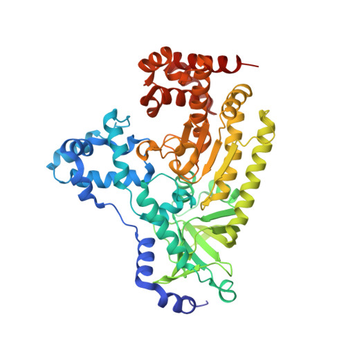
Anthrax toxin lethal factor domain 3 is highly mobile and responsive to ligand binding.

Maize, K.M.Kurbanov, E.K.De La Mora-Rey, T.Geders, T.W.Hwang, D.J.Walters, M.A.Johnson, R.L.Amin, E.A.Finzel, B.C.

(2014) Acta Crystallogr D Biol Crystallogr 70: 2813-2822

  • DOI: https://doi.org/10.1107/S1399004714018161
  • Primary Citation of Related Structures:  
    4PKQ, 4PKR, 4PKS, 4PKT, 4PKU, 4PKV, 4PKW

  • PubMed Abstract: 

    The secreted anthrax toxin consists of three components: the protective antigen (PA), edema factor (EF) and lethal factor (LF). LF, a zinc metalloproteinase, compromises the host immune system primarily by targeting mitogen-activated protein kinase kinases in macrophages. Peptide substrates and small-molecule inhibitors bind LF in the space between domains 3 and 4 of the hydrolase. Domain 3 is attached on a hinge to domain 2 via residues Ile300 and Pro385, and can move through an angular arc of greater than 35° in response to the binding of different ligands. Here, multiple LF structures including five new complexes with co-crystallized inhibitors are compared and three frequently populated LF conformational states termed `bioactive', `open' and `tight' are identified. The bioactive position is observed with large substrate peptides and leaves all peptide-recognition subsites open and accessible. The tight state is seen in unliganded and small-molecule complex structures. In this state, domain 3 is clamped over certain substrate subsites, blocking access. The open position appears to be an intermediate state between these extremes and is observed owing to steric constraints imposed by specific bound ligands. The tight conformation may be the lowest-energy conformation among the reported structures, as it is the position observed with no bound ligand, while the open and bioactive conformations are likely to be ligand-induced.


  • Organizational Affiliation

    Department of Medicinal Chemistry and Minnesota Supercomputing Institute, University of Minnesota, 8-101 Weaver-Densford Hall, 308 Harvard Street SE, Minneapolis, MN 55455, USA.


Macromolecules
Find similar proteins by:  (by identity cutoff)  |  3D Structure
Entity ID: 1
MoleculeChains Sequence LengthOrganismDetailsImage
Lethal factor519Bacillus anthracisMutation(s): 1 
Gene Names: lefpXO1-107BXA0172GBAA_pXO1_0172
EC: 3.4.24.83
UniProt
Find proteins for P15917 (Bacillus anthracis)
Explore P15917 
Go to UniProtKB:  P15917
Entity Groups  
Sequence Clusters30% Identity50% Identity70% Identity90% Identity95% Identity100% Identity
UniProt GroupP15917
Sequence Annotations
Expand
  • Reference Sequence
Small Molecules
Ligands 2 Unique
IDChains Name / Formula / InChI Key2D Diagram3D Interactions
30R
Query on 30R

Download Ideal Coordinates CCD File 
B [auth A]N~2~-[4-(aminomethyl)benzyl]-N~2~-[(4-fluoro-3-methylphenyl)sulfonyl]-N-hydroxy-D-alaninamide
C18 H22 F N3 O4 S
CWTKLEFNKQTFGU-CYBMUJFWSA-N
ZN
Query on ZN

Download Ideal Coordinates CCD File 
C [auth A]ZINC ION
Zn
PTFCDOFLOPIGGS-UHFFFAOYSA-N
Binding Affinity Annotations 
IDSourceBinding Affinity
30R Binding MOAD:  4PKV IC50: 5600 (nM) from 1 assay(s)
Experimental Data & Validation

Experimental Data

  • Method: X-RAY DIFFRACTION
  • Resolution: 2.50 Å
  • R-Value Free: 0.253 
  • R-Value Work: 0.181 
  • R-Value Observed: 0.185 
  • Space Group: P 21 21 21
Unit Cell:
Length ( Å )Angle ( ˚ )
a = 57.48α = 90
b = 78.729β = 90
c = 139.09γ = 90
Software Package:
Software NamePurpose
XDSdata reduction
SCALAdata scaling
PHASERphasing
PHENIXrefinement
PDB_EXTRACTdata extraction

Structure Validation

View Full Validation Report


Ligand Structure Quality Assessment 


Entry History & Funding Information

Deposition Data


Funding OrganizationLocationGrant Number
Minnesota Dept. of Employment and Economic DevelopmentUnited StatesSPAP-06-0014-P-FY07
National Institutes of Health/National Institute of General Medical Sciences (NIH/NIGMS)United StatesGM08700
National Institutes of Health/National Institute Of Allergy and Infectious Diseases (NIH/NIAID)United StatesAI091790-01

Revision History  (Full details and data files)

  • Version 1.0: 2014-11-12
    Type: Initial release
  • Version 1.1: 2014-11-26
    Changes: Database references
  • Version 1.2: 2015-02-04
    Changes: Derived calculations
  • Version 1.3: 2017-09-27
    Changes: Author supporting evidence, Derived calculations, Other, Refinement description, Source and taxonomy, Structure summary
  • Version 1.4: 2019-12-11
    Changes: Author supporting evidence
  • Version 1.5: 2023-12-27
    Changes: Data collection, Database references, Refinement description