4PKE

The structure of a conserved Piezo channel domain reveals a novel beta sandwich fold


Experimental Data Snapshot

  • Method: X-RAY DIFFRACTION
  • Resolution: 2.50 Å
  • R-Value Free: 0.238 
  • R-Value Work: 0.228 
  • R-Value Observed: 0.229 

wwPDB Validation   3D Report Full Report


This is version 1.7 of the entry. See complete history


Literature

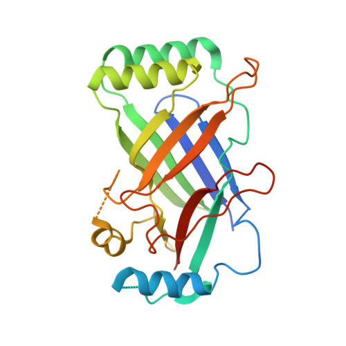
The Structure of a Conserved Piezo Channel Domain Reveals a Topologically Distinct beta Sandwich Fold.

Kamajaya, A.Kaiser, J.T.Lee, J.Reid, M.Rees, D.C.

(2014) Structure 22: 1520-1527

  • DOI: https://doi.org/10.1016/j.str.2014.08.009
  • Primary Citation of Related Structures:  
    4PKE, 4PKX

  • PubMed Abstract: 

    Piezo has recently been identified as a family of eukaryotic mechanosensitive channels composed of subunits containing over 2,000 amino acids, without recognizable sequence similarity to other channels. Here, we present the crystal structure of a large, conserved extramembrane domain located just before the last predicted transmembrane helix of C. elegans PIEZO, which adopts a topologically distinct β sandwich fold. The structure was also determined of a point mutation located on a conserved surface at the position equivalent to the human PIEZO1 mutation found in dehydrated hereditary stomatocytosis patients (M2225R). While the point mutation does not change the overall domain structure, it does alter the surface electrostatic potential that may perturb interactions with a yet-to-be-identified ligand or protein. The lack of structural similarity between this domain and any previously characterized fold, including those of eukaryotic and bacterial channels, highlights the distinctive nature of the Piezo family of eukaryotic mechanosensitive channels.


  • Organizational Affiliation

    Biochemistry and Molecular Biophysics Graduate Option, California Institute of Technology, Pasadena, CA 91125, USA; Division of Chemistry and Chemical Engineering, California Institute of Technology, Pasadena, CA 91125, USA.


Macromolecules
Find similar proteins by:  (by identity cutoff)  |  3D Structure
Entity ID: 1
MoleculeChains Sequence LengthOrganismDetailsImage
Protein C10C5.1, isoform i291Caenorhabditis elegansMutation(s): 0 
Gene Names: C10C5.1CELE_C10C5.1T20D3.11
UniProt
Find proteins for A0A061ACU2 (Caenorhabditis elegans)
Explore A0A061ACU2 
Go to UniProtKB:  A0A061ACU2
Entity Groups  
Sequence Clusters30% Identity50% Identity70% Identity90% Identity95% Identity100% Identity
UniProt GroupA0A061ACU2
Sequence Annotations
Expand
  • Reference Sequence
Experimental Data & Validation

Experimental Data

  • Method: X-RAY DIFFRACTION
  • Resolution: 2.50 Å
  • R-Value Free: 0.238 
  • R-Value Work: 0.228 
  • R-Value Observed: 0.229 
  • Space Group: H 3 2
Unit Cell:
Length ( Å )Angle ( ˚ )
a = 72.588α = 90
b = 72.588β = 90
c = 241.551γ = 120
Software Package:
Software NamePurpose
PHENIXrefinement

Structure Validation

View Full Validation Report


Entry History & Funding Information

Deposition Data


Funding OrganizationLocationGrant Number
Howard Hughes Medical Institute (HHMI)United States--

Revision History  (Full details and data files)

  • Version 1.0: 2014-10-08
    Type: Initial release
  • Version 1.1: 2014-10-22
    Changes: Database references
  • Version 1.2: 2014-10-29
    Changes: Atomic model, Derived calculations
  • Version 1.3: 2014-11-05
    Changes: Database references
  • Version 1.4: 2016-07-27
    Changes: Data collection
  • Version 1.5: 2017-09-13
    Changes: Author supporting evidence, Derived calculations
  • Version 1.6: 2019-11-20
    Changes: Author supporting evidence
  • Version 1.7: 2023-12-27
    Changes: Data collection, Database references, Derived calculations