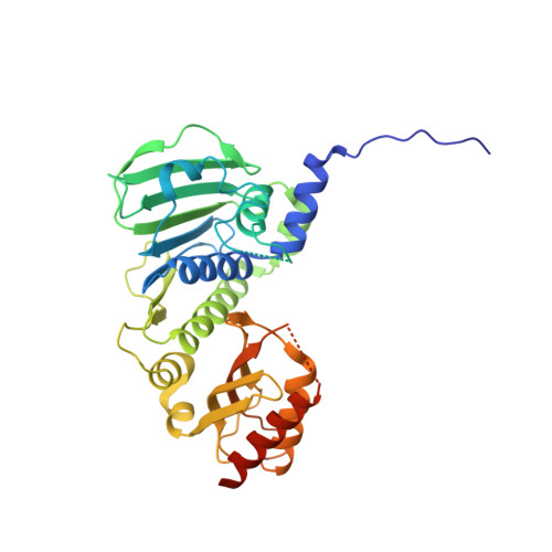
4P7A

Crystal Structure of human MLH1


Experimental Data Snapshot

  • Method: X-RAY DIFFRACTION
  • Resolution: 2.30 Å
  • R-Value Free: 0.254 
  • R-Value Work: 0.203 
  • R-Value Observed: 0.205 

wwPDB Validation   3D Report Full Report


Ligand Structure Quality Assessment 


This is version 2.1 of the entry. See complete history

Re-refinement Note

This entry reflects an alternative modeling of the original data in: 3NA3


Literature

Structure of the human MLH1 N-terminus: implications for predisposition to Lynch syndrome.

Wu, H.Zeng, H.Lam, R.Tempel, W.Kerr, I.D.Min, J.

(2015) Acta Crystallogr F Struct Biol Commun 71: 981-985

  • DOI: https://doi.org/10.1107/S2053230X15010183
  • Primary Citation of Related Structures:  
    4P7A

  • PubMed Abstract: 

    Mismatch repair prevents the accumulation of erroneous insertions/deletions and non-Watson-Crick base pairs in the genome. Pathogenic mutations in the MLH1 gene are associated with a predisposition to Lynch and Turcot's syndromes. Although genetic testing for these mutations is available, robust classification of variants requires strong clinical and functional support. Here, the first structure of the N-terminus of human MLH1, determined by X-ray crystallography, is described. The structure shares a high degree of similarity with previously determined prokaryotic MLH1 homologs; however, this structure affords a more accurate platform for the classification of MLH1 variants.


  • Organizational Affiliation

    Structural Genomics Consortium, University of Toronto, 101 College Street, Toronto, ON M5G 1L7, Canada.


Macromolecules
Find similar proteins by:  (by identity cutoff)  |  3D Structure
Entity ID: 1
MoleculeChains Sequence LengthOrganismDetailsImage
DNA mismatch repair protein Mlh1348Homo sapiensMutation(s): 0 
Gene Names: MLH1COCA2
UniProt & NIH Common Fund Data Resources
Find proteins for P40692 (Homo sapiens)
Explore P40692 
Go to UniProtKB:  P40692
PHAROS:  P40692
GTEx:  ENSG00000076242 
Entity Groups  
Sequence Clusters30% Identity50% Identity70% Identity90% Identity95% Identity100% Identity
UniProt GroupP40692
Sequence Annotations
Expand
  • Reference Sequence
Small Molecules
Ligands 3 Unique
IDChains Name / Formula / InChI Key2D Diagram3D Interactions
ADP
Query on ADP

Download Ideal Coordinates CCD File 
C [auth A]ADENOSINE-5'-DIPHOSPHATE
C10 H15 N5 O10 P2
XTWYTFMLZFPYCI-KQYNXXCUSA-N
MG
Query on MG

Download Ideal Coordinates CCD File 
B [auth A]MAGNESIUM ION
Mg
JLVVSXFLKOJNIY-UHFFFAOYSA-N
UNX
Query on UNX

Download Ideal Coordinates CCD File 
D [auth A]
E [auth A]
F [auth A]
G [auth A]
H [auth A]
D [auth A],
E [auth A],
F [auth A],
G [auth A],
H [auth A],
I [auth A],
J [auth A],
K [auth A],
L [auth A]
UNKNOWN ATOM OR ION
X
Experimental Data & Validation

Experimental Data

  • Method: X-RAY DIFFRACTION
  • Resolution: 2.30 Å
  • R-Value Free: 0.254 
  • R-Value Work: 0.203 
  • R-Value Observed: 0.205 
  • Space Group: P 64
Unit Cell:
Length ( Å )Angle ( ˚ )
a = 94.568α = 90
b = 94.568β = 90
c = 85.825γ = 120
Software Package:
Software NamePurpose
REFMACrefinement
Aimlessdata scaling
BUSTERrefinement
XDSdata reduction
PDB_EXTRACTdata extraction

Structure Validation

View Full Validation Report


Ligand Structure Quality Assessment 


Entry History 

Deposition Data

Revision History  (Full details and data files)

  • Version 1.0: 2014-04-09
    Type: Initial release
  • Version 1.1: 2014-11-12
    Changes: Other
  • Version 1.2: 2015-12-16
    Changes: Database references
  • Version 2.0: 2019-07-17
    Changes: Advisory, Atomic model, Data collection, Derived calculations, Refinement description, Structure summary
  • Version 2.1: 2023-12-27
    Changes: Data collection, Database references, Derived calculations