4P3I

Structure of the P domain from a GI.7 Norovirus variant in complex with LeA HBGA.


Experimental Data Snapshot

  • Method: X-RAY DIFFRACTION
  • Resolution: 1.69 Å
  • R-Value Free: 0.177 
  • R-Value Work: 0.155 
  • R-Value Observed: 0.156 

wwPDB Validation   3D Report Full Report


This is version 3.2 of the entry. See complete history


Literature

Structural analysis of determinants of histo-blood group antigen binding specificity in genogroup I noroviruses.

Shanker, S.Czako, R.Sankaran, B.Atmar, R.L.Estes, M.K.Prasad, B.V.

(2014) J Virol 88: 6168-6180

  • DOI: https://doi.org/10.1128/JVI.00201-14
  • Primary Citation of Related Structures:  
    4P12, 4P1V, 4P25, 4P26, 4P2N, 4P3I

  • PubMed Abstract: 

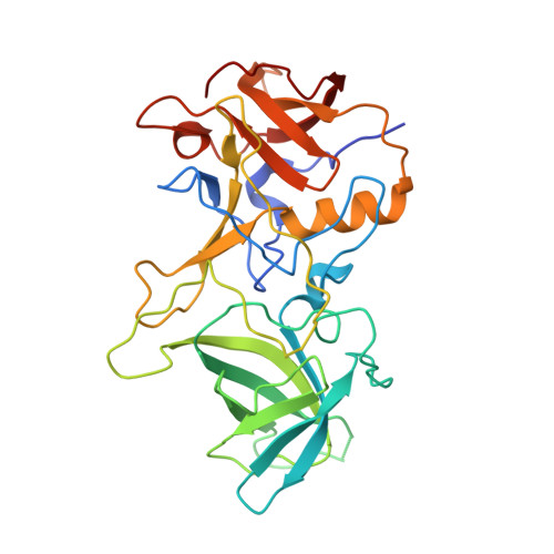
    Human noroviruses (NoVs) cause acute epidemic gastroenteritis. Susceptibility to the majority of NoV infections is determined by genetically controlled secretor-dependent expression of histo-blood group antigens (HBGAs), which are also critical for NoV attachment to host cells. Human NoVs are classified into two major genogroups (genogroup I [GI] and GII), with each genogroup further divided into several genotypes. GII NoVs are more prevalent and exhibit periodic emergence of new variants, suggested to be driven by altered HBGA binding specificities and antigenic drift. Recent epidemiological studies show increased activity among GI NoVs, with some members showing the ability to bind nonsecretor HBGAs. NoVs bind HBGAs through the protruding (P) domain of the major capsid protein VP1. GI NoVs, similar to GII, exhibit significant sequence variations in the P domain; it is unclear how these variations affect HBGA binding specificities. To understand the determinants of possible strain-specific HBGA binding among GI NoVs, we determined the structure of the P domain of a GI.7 clinical isolate and compared it to the previously determined P domain structures of GI.1 and GI.2 strains. Our crystallographic studies revealed significant structural differences, particularly in the loop regions of the GI.7 P domain, altering its surface topography and electrostatic landscape and potentially indicating antigenic variation. The GI.7 strain bound to H- and A-type, Lewis secretor, and Lewis nonsecretor families of HBGAs, allowing us to further elucidate the structural determinants of nonsecretor HBGA binding among GI NoVs and to infer several contrasting and generalizable features of HBGA binding in the GI NoVs.


  • Organizational Affiliation

    Verna Marrs McLean Department of Biochemistry and Molecular Biology, Baylor College of Medicine, Houston, Texas, USA.


Macromolecules
Find similar proteins by:  (by identity cutoff)  |  3D Structure
Entity ID: 1
MoleculeChains Sequence LengthOrganismDetailsImage
P domain of VP1
A, B, C, D
301Norovirus Hu/GI.7/TCH-060/USA/2003Mutation(s): 0 
UniProt
Find proteins for G8FL04 (Norovirus Hu/GI.7/TCH-060/USA/2003)
Explore G8FL04 
Go to UniProtKB:  G8FL04
Entity Groups  
Sequence Clusters30% Identity50% Identity70% Identity90% Identity95% Identity100% Identity
UniProt GroupG8FL04
Sequence Annotations
Expand
  • Reference Sequence
Oligosaccharides

Help

Entity ID: 2
MoleculeChains Length2D Diagram Glycosylation3D Interactions
beta-D-galactopyranose-(1-3)-[alpha-L-fucopyranose-(1-4)]2-acetamido-2-deoxy-beta-D-glucopyranose
E, F, G, H
3N/A
Glycosylation Resources
GlyTouCan:  G39023AU
GlyCosmos:  G39023AU
GlyGen:  G39023AU
Experimental Data & Validation

Experimental Data

  • Method: X-RAY DIFFRACTION
  • Resolution: 1.69 Å
  • R-Value Free: 0.177 
  • R-Value Work: 0.155 
  • R-Value Observed: 0.156 
  • Space Group: P 1
Unit Cell:
Length ( Å )Angle ( ˚ )
a = 62.168α = 72.77
b = 63.277β = 82.2
c = 90.4γ = 60.31
Software Package:
Software NamePurpose
PHENIXrefinement

Structure Validation

View Full Validation Report


Entry History & Funding Information

Deposition Data


Funding OrganizationLocationGrant Number
National Institutes of Health/National Institute Of Allergy and Infectious Diseases (NIH/NIAID)United States--
National Institutes of Health/National Institute of Diabetes and Digestive and Kidney Disease (NIH/NIDDK)United States--
National Institutes of Health/National Cancer Institute (NIH/NCI)United States--

Revision History  (Full details and data files)

  • Version 1.0: 2014-04-02
    Type: Initial release
  • Version 1.1: 2014-05-21
    Changes: Database references
  • Version 2.0: 2018-03-21
    Changes: Atomic model, Data collection, Database references, Derived calculations, Other, Source and taxonomy, Structure summary
  • Version 2.1: 2019-11-27
    Changes: Author supporting evidence, Data collection
  • Version 3.0: 2020-07-29
    Type: Remediation
    Reason: Carbohydrate remediation
    Changes: Atomic model, Data collection, Derived calculations, Refinement description, Structure summary
  • Version 3.1: 2020-08-05
    Changes: Structure summary
  • Version 3.2: 2023-12-27
    Changes: Data collection, Database references, Structure summary