4P1M

The structure of Escherichia coli ZapA


Experimental Data Snapshot

  • Method: X-RAY DIFFRACTION
  • Resolution: 1.95 Å
  • R-Value Free: 0.270 
  • R-Value Work: 0.224 
  • R-Value Observed: 0.226 

wwPDB Validation   3D Report Full Report


This is version 1.4 of the entry. See complete history


Literature

Crystal Structure and Site-directed Mutational Analysis Reveals Key Residues Involved in Escherichia coli ZapA Function.

Roach, E.J.Kimber, M.S.Khursigara, C.M.

(2014) J Biol Chem 289: 23276-23286

  • DOI: https://doi.org/10.1074/jbc.M114.561928
  • Primary Citation of Related Structures:  
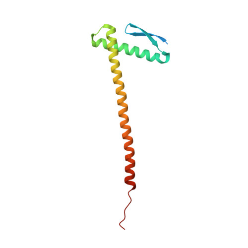
    4P1M

  • PubMed Abstract: 

    FtsZ is an essential cell division protein in Escherichia coli, and its localization, filamentation, and bundling at the mid-cell are required for Z-ring stability. Once assembled, the Z-ring recruits a series of proteins that comprise the bacterial divisome. Zaps (FtsZ-associated proteins) stabilize the Z-ring by increasing lateral interactions between individual filaments, bundling FtsZ to provide a scaffold for divisome assembly. The x-ray crystallographic structure of E. coli ZapA was determined, identifying key structural differences from the existing ZapA structure from Pseudomonas aeruginosa, including a charged α-helix on the globular domains of the ZapA tetramer. Key helix residues in E. coli ZapA were modified using site-directed mutagenesis. These ZapA variants significantly decreased FtsZ bundling in protein sedimentation assays when compared with WT ZapA proteins. Electron micrographs of ZapA-bundled FtsZ filaments showed the modified ZapA variants altered the number of FtsZ filaments per bundle. These in vitro results were corroborated in vivo by expressing the ZapA variants in an E. coli ΔzapA strain. In vivo, ZapA variants that altered FtsZ bundling showed an elongated phenotype, indicating improper cell division. Our findings highlight the importance of key ZapA residues that influence the extent of FtsZ bundling and that ultimately affect Z-ring formation in dividing cells.


  • Organizational Affiliation

    From the Department of Molecular and Cellular Biology, University of Guelph, Guelph, Ontario N1G 2W1, Canada.


Macromolecules
Find similar proteins by:  (by identity cutoff)  |  3D Structure
Entity ID: 1
MoleculeChains Sequence LengthOrganismDetailsImage
Cell division protein ZapA
A, B
123Escherichia coliMutation(s): 0 
Gene Names: zapA
UniProt
Find proteins for P0ADS2 (Escherichia coli (strain K12))
Explore P0ADS2 
Go to UniProtKB:  P0ADS2
Entity Groups  
Sequence Clusters30% Identity50% Identity70% Identity90% Identity95% Identity100% Identity
UniProt GroupP0ADS2
Sequence Annotations
Expand
  • Reference Sequence
Small Molecules
Ligands 1 Unique
IDChains Name / Formula / InChI Key2D Diagram3D Interactions
CL
Query on CL

Download Ideal Coordinates CCD File 
C [auth B]CHLORIDE ION
Cl
VEXZGXHMUGYJMC-UHFFFAOYSA-M
Experimental Data & Validation

Experimental Data

  • Method: X-RAY DIFFRACTION
  • Resolution: 1.95 Å
  • R-Value Free: 0.270 
  • R-Value Work: 0.224 
  • R-Value Observed: 0.226 
  • Space Group: P 61 2 2
Unit Cell:
Length ( Å )Angle ( ˚ )
a = 54.1α = 90
b = 54.1β = 90
c = 328.07γ = 120
Software Package:
Software NamePurpose
PHENIXrefinement

Structure Validation

View Full Validation Report


Entry History & Funding Information

Deposition Data


Funding OrganizationLocationGrant Number
Natural Sciences and Engineering Research Council (NSERC, Canada)Canada327280
Natural Sciences and Engineering Research Council (NSERC, Canada)Canada371639

Revision History  (Full details and data files)

  • Version 1.0: 2014-07-16
    Type: Initial release
  • Version 1.1: 2014-10-01
    Changes: Database references
  • Version 1.2: 2017-09-20
    Changes: Advisory, Author supporting evidence, Database references, Derived calculations, Other, Source and taxonomy
  • Version 1.3: 2020-01-08
    Changes: Author supporting evidence
  • Version 1.4: 2023-09-27
    Changes: Data collection, Database references, Refinement description