Structure of the type VI secretion phospholipase effector Tle1 provides insight into its hydrolysis and membrane targeting.
Hu, H.D., Zhang, H., Gao, Z.Q., Wang, D., Liu, G., Xu, J., Lan, K., Dong, Y.H.(2014) Acta Crystallogr D Biol Crystallogr 70: 2175-2185
- PubMed: 25084336
- DOI: https://doi.org/10.1107/S1399004714012899
- Primary Citation of Related Structures:
4O5P - PubMed Abstract:
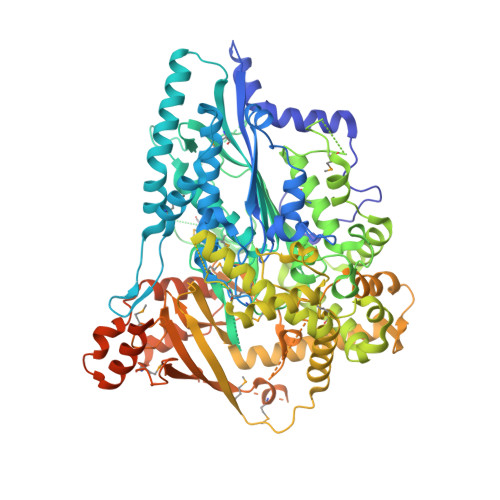
A diverse superfamily of phospholipases consisting of the type VI lipase effectors Tle1-Tle5 secreted by the bacterial type VI secretion system (T6SS) have recently been identified as antibacterial effectors that hydrolyze membrane phospholipids. These effectors show no significant homology to known lipases, and their mechanism of membrane targeting and hydrolysis of phospholipids remains unknown. Here, the crystal structure of Tle1 (∼96.5 kDa) from Pseudomonas aeruginosa refined to 2.0 Å resolution is reported, representing the first structure of this superfamily. Its overall structure can be divided into two distinct parts, the phospholipase catalytic module and the putative membrane-anchoring module; this arrangement has not previously been observed in known lipase structures. The phospholipase catalytic module has a canonical α/β-hydrolase fold and mutation of any residue in the Ser-Asp-His catalytic triad abolishes its toxicity. The putative membrane-anchoring module adopts an open conformation composed of three amphipathic domains, and its partial folds are similar to those of several periplasmic or membrane proteins. A cell-toxicity assay revealed that the putative membrane-anchoring module is critical to Tle1 antibacterial activity. A molecular-dynamics (MD) simulation system in which the putative membrane-anchoring module embedded into a bilayer was stable over 50 ns. These structure-function studies provide insight into the hydrolysis and membrane-targeting process of the unique phospholipase Tle1.
Organizational Affiliation:
Key Laboratory of Molecular Virology and Immunology, Institute Pasteur of Shanghai, Chinese Academy of Sciences, Shanghai 200025, People's Republic of China.