4NZI

Crystal structure of murine neuroglobin mutant V140W


Experimental Data Snapshot

  • Method: X-RAY DIFFRACTION
  • Resolution: 2.10 Å
  • R-Value Free: 0.262 
  • R-Value Work: 0.219 
  • R-Value Observed: 0.223 

wwPDB Validation   3D Report Full Report


Ligand Structure Quality Assessment 


This is version 1.2 of the entry. See complete history


Literature

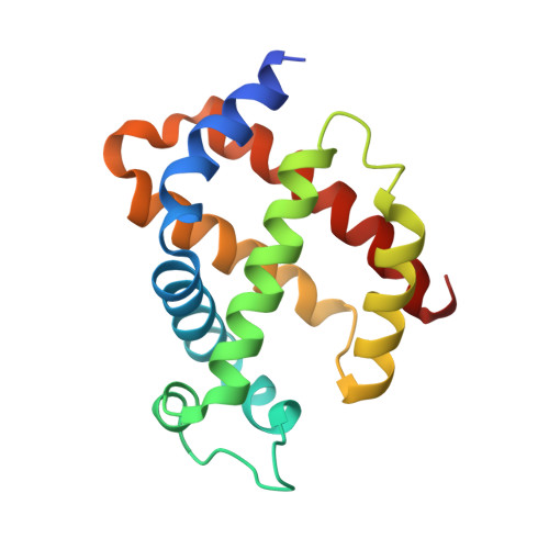
Engineering the internal cavity of neuroglobin demonstrates the role of the haem-sliding mechanism.

Avella, G.Ardiccioni, C.Scaglione, A.Moschetti, T.Rondinelli, C.Montemiglio, L.C.Savino, C.Giuffre, A.Brunori, M.Vallone, B.

(2014) Acta Crystallogr D Biol Crystallogr 70: 1640-1648

  • DOI: https://doi.org/10.1107/S1399004714007032
  • Primary Citation of Related Structures:  
    4MU5, 4NZI, 4O1T, 4O2G, 4O35

  • PubMed Abstract: 

    Neuroglobin is a member of the globin family involved in neuroprotection; it is primarily expressed in the brain and retina of vertebrates. Neuroglobin belongs to the heterogeneous group of hexacoordinate globins that have evolved in animals, plants and bacteria, endowed with the capability of reversible intramolecular coordination, allowing the binding of small gaseous ligands (O2, NO and CO). In a unique fashion among haemoproteins, ligand-binding events in neuroglobin are dependent on the sliding of the haem itself within a preformed internal cavity, as revealed by the crystal structure of its CO-bound derivative. Point mutants of the neuroglobin internal cavity have been engineered and their functional and structural characterization shows that hindering the haem displacement leads to a decrease in CO affinity, whereas reducing the cavity volume without interfering with haem sliding has negligible functional effects.


  • Organizational Affiliation

    Istituto Pasteur-Fondazione Cenci Bolognetti and Istituto di Biologia e Patologia Molecolari del CNR, Dipartimento di Scienze Biochimiche `A. Rossi Fanelli', Sapienza Università di Roma, Piazzale A. Moro 5, 00185 Rome, Italy.


Macromolecules
Find similar proteins by:  (by identity cutoff)  |  3D Structure
Entity ID: 1
MoleculeChains Sequence LengthOrganismDetailsImage
Neuroglobin154Mus musculusMutation(s): 3 
Gene Names: Ngb
UniProt
Find proteins for Q9ER97 (Mus musculus)
Explore Q9ER97 
Go to UniProtKB:  Q9ER97
Entity Groups  
Sequence Clusters30% Identity50% Identity70% Identity90% Identity95% Identity100% Identity
UniProt GroupQ9ER97
Sequence Annotations
Expand
  • Reference Sequence
Small Molecules
Ligands 2 Unique
IDChains Name / Formula / InChI Key2D Diagram3D Interactions
HEM
Query on HEM

Download Ideal Coordinates CCD File 
B [auth A]PROTOPORPHYRIN IX CONTAINING FE
C34 H32 Fe N4 O4
KABFMIBPWCXCRK-RGGAHWMASA-L
SO4
Query on SO4

Download Ideal Coordinates CCD File 
C [auth A]SULFATE ION
O4 S
QAOWNCQODCNURD-UHFFFAOYSA-L
Experimental Data & Validation

Experimental Data

  • Method: X-RAY DIFFRACTION
  • Resolution: 2.10 Å
  • R-Value Free: 0.262 
  • R-Value Work: 0.219 
  • R-Value Observed: 0.223 
  • Space Group: H 3 2
Unit Cell:
Length ( Å )Angle ( ˚ )
a = 87.214α = 90
b = 87.214β = 90
c = 114.399γ = 120
Software Package:
Software NamePurpose
PHENIXrefinement
PDB_EXTRACTdata extraction
DNAdata collection
MOSFLMdata reduction
SCALAdata scaling
PHASERphasing

Structure Validation

View Full Validation Report


Ligand Structure Quality Assessment 


Entry History 

Deposition Data

Revision History  (Full details and data files)

  • Version 1.0: 2014-06-18
    Type: Initial release
  • Version 1.1: 2014-09-24
    Changes: Database references
  • Version 1.2: 2024-02-28
    Changes: Data collection, Database references, Derived calculations