4NSE

BOVINE ENDOTHELIAL NITRIC OXIDE SYNTHASE, H4B-FREE, L-ARG COMPLEX


Experimental Data Snapshot

  • Method: X-RAY DIFFRACTION
  • Resolution: 1.95 Å
  • R-Value Free: 0.260 
  • R-Value Work: 0.212 
  • R-Value Observed: 0.212 

wwPDB Validation   3D Report Full Report


This is version 1.3 of the entry. See complete history


Literature

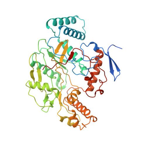
Crystal structure of constitutive endothelial nitric oxide synthase: a paradigm for pterin function involving a novel metal center.

Raman, C.S.Li, H.Martasek, P.Kral, V.Masters, B.S.Poulos, T.L.

(1998) Cell 95: 939-950

  • DOI: https://doi.org/10.1016/s0092-8674(00)81718-3
  • Primary Citation of Related Structures:  
    1NSE, 2NSE, 3NSE, 4NSE

  • PubMed Abstract: 

    Nitric oxide, a key signaling molecule, is produced by a family of enzymes collectively called nitric oxide synthases (NOS). Here, we report the crystal structure of the heme domain of endothelial NOS in tetrahydrobiopterin (H4B)-free and -bound forms at 1.95 A and 1.9 A resolution, respectively. In both structures a zinc ion is tetrahedrally coordinated to pairs of symmetry-related cysteine residues at the dimer interface. The phylogenetically conserved Cys-(X)4-Cys motif and its strategic location establish a structural role for the metal center in maintaining the integrity of the H4B-binding site. The unexpected recognition of the substrate, L-arginine, at the H4B site indicates that this site is poised to stabilize a positively charged pterin ring and suggests a model involving a cationic pterin radical in the catalytic cycle.


  • Organizational Affiliation

    Department of Molecular Biology and Biochemistry, University of California, Irvine 92697-3900, USA.


Macromolecules
Find similar proteins by:  (by identity cutoff)  |  3D Structure
Entity ID: 1
MoleculeChains Sequence LengthOrganismDetailsImage
NITRIC OXIDE SYNTHASE
A, B
444Bos taurusMutation(s): 0 
EC: 1.14.13.39
UniProt
Find proteins for P29473 (Bos taurus)
Explore P29473 
Go to UniProtKB:  P29473
Entity Groups  
Sequence Clusters30% Identity50% Identity70% Identity90% Identity95% Identity100% Identity
UniProt GroupP29473
Sequence Annotations
Expand
  • Reference Sequence
Small Molecules
Ligands 6 Unique
IDChains Name / Formula / InChI Key2D Diagram3D Interactions
HEM
Query on HEM

Download Ideal Coordinates CCD File 
F [auth A],
M [auth B]
PROTOPORPHYRIN IX CONTAINING FE
C34 H32 Fe N4 O4
KABFMIBPWCXCRK-RGGAHWMASA-L
ARG
Query on ARG

Download Ideal Coordinates CCD File 
E [auth A],
L [auth B]
ARGININE
C6 H15 N4 O2
ODKSFYDXXFIFQN-BYPYZUCNSA-O
CAC
Query on CAC

Download Ideal Coordinates CCD File 
D [auth A],
J [auth B]
CACODYLATE ION
C2 H6 As O2
OGGXGZAMXPVRFZ-UHFFFAOYSA-M
GOL
Query on GOL

Download Ideal Coordinates CCD File 
G [auth A],
H [auth A],
N [auth B]
GLYCEROL
C3 H8 O3
PEDCQBHIVMGVHV-UHFFFAOYSA-N
ZN
Query on ZN

Download Ideal Coordinates CCD File 
K [auth B]ZINC ION
Zn
PTFCDOFLOPIGGS-UHFFFAOYSA-N
ACT
Query on ACT

Download Ideal Coordinates CCD File 
C [auth A],
I [auth B]
ACETATE ION
C2 H3 O2
QTBSBXVTEAMEQO-UHFFFAOYSA-M
Experimental Data & Validation

Experimental Data

  • Method: X-RAY DIFFRACTION
  • Resolution: 1.95 Å
  • R-Value Free: 0.260 
  • R-Value Work: 0.212 
  • R-Value Observed: 0.212 
  • Space Group: P 21 21 21
Unit Cell:
Length ( Å )Angle ( ˚ )
a = 57.66α = 90
b = 106.1β = 90
c = 155.75γ = 90
Software Package:
Software NamePurpose
DENZOdata reduction
SCALEPACKdata scaling
X-PLORmodel building
X-PLORrefinement
X-PLORphasing

Structure Validation

View Full Validation Report


Entry History 

Deposition Data

Revision History  (Full details and data files)

  • Version 1.0: 1999-05-18
    Type: Initial release
  • Version 1.1: 2008-03-25
    Changes: Version format compliance
  • Version 1.2: 2011-07-13
    Changes: Non-polymer description, Version format compliance
  • Version 1.3: 2024-02-28
    Changes: Data collection, Database references, Derived calculations