Dimerization of VirD2 Binding Protein Is Essential for Agrobacterium Induced Tumor Formation in Plants
Padavannil, A., Jobichen, C., Qinghua, Y., Seetharaman, J., Velazquez-campoy, A., Yang, L., Pan, S.Q., Sivaraman, J.(2014) PLoS Pathog 10: e1003948-e1003948
- PubMed: 24626239
- DOI: https://doi.org/10.1371/journal.ppat.1003948
- Primary Citation of Related Structures:
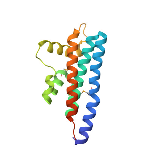
4NQF - PubMed Abstract:
The Type IV Secretion System (T4SS) is the only bacterial secretion system known to translocate both DNA and protein substrates. The VirB/D4 system from Agrobacterium tumefaciens is a typical T4SS. It facilitates the bacteria to translocate the VirD2-T-DNA complex to the host cell cytoplasm. In addition to protein-DNA complexes, the VirB/D4 system is also involved in the translocation of several effector proteins, including VirE2, VirE3 and VirF into the host cell cytoplasm. These effector proteins aid in the proper integration of the translocated DNA into the host genome. The VirD2-binding protein (VBP) is a key cytoplasmic protein that recruits the VirD2-T-DNA complex to the VirD4-coupling protein (VirD4 CP) of the VirB/D4 T4SS apparatus. Here, we report the crystal structure and associated functional studies of the C-terminal domain of VBP. This domain mainly consists of α-helices, and the two monomers of the asymmetric unit form a tight dimer. The structural analysis of this domain confirms the presence of a HEPN (higher eukaryotes and prokaryotes nucleotide-binding) fold. Biophysical studies show that VBP is a dimer in solution and that the HEPN domain is the dimerization domain. Based on structural and mutagenesis analyses, we show that substitution of key residues at the interface disrupts the dimerization of both the HEPN domain and full-length VBP. In addition, pull-down analyses show that only dimeric VBP can interact with VirD2 and VirD4 CP. Finally, we show that only Agrobacterium harboring dimeric full-length VBP can induce tumors in plants. This study sheds light on the structural basis of the substrate recruiting function of VBP in the T4SS pathway of A. tumefaciens and in other pathogenic bacteria employing similar systems.
Organizational Affiliation:
Department of Biological Sciences, National University of Singapore, Singapore.