


Crystal structure of 5-carboxyvanillate decarboxylase LigW from Sphingomonas paucimobilis complexed with Mn and 5-methoxyisophtalic acid
Fedorov, A.A., Fedorov, E.V., Vladimirova, A., Raushel, F.M., Almo, S.C.To be published.
Experimental Data Snapshot
Entity ID: 1 | |||||
|---|---|---|---|---|---|
| Molecule | Chains | Sequence Length | Organism | Details | Image |
| 5-carboxyvanillate decarboxylase | 335 | Sphingomonas paucimobilis | Mutation(s): 0 Gene Names: ligW | 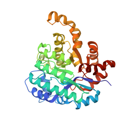 | |
UniProt | |||||
Find proteins for Q8RJ47 (Sphingomonas paucimobilis) Explore Q8RJ47 Go to UniProtKB: Q8RJ47 | |||||
Entity Groups | |||||
| Sequence Clusters | 30% Identity50% Identity70% Identity90% Identity95% Identity100% Identity | ||||
| UniProt Group | Q8RJ47 | ||||
Sequence AnnotationsExpand | |||||
| |||||
| Ligands 4 Unique | |||||
|---|---|---|---|---|---|
| ID | Chains | Name / Formula / InChI Key | 2D Diagram | 3D Interactions | |
| 1WB Query on 1WB | AA [auth E] DA [auth F] J [auth A] KA [auth G] P [auth B] | 5-methoxybenzene-1,3-dicarboxylic acid C9 H8 O5 POSMIIJADZKUPL-UHFFFAOYSA-N |  | ||
| GOL Query on GOL | BA [auth E], EA [auth F] | GLYCEROL C3 H8 O3 PEDCQBHIVMGVHV-UHFFFAOYSA-N |  | ||
| EDO Query on EDO | FA [auth F] GA [auth F] HA [auth F] IA [auth F] K [auth A] | 1,2-ETHANEDIOL C2 H6 O2 LYCAIKOWRPUZTN-UHFFFAOYSA-N |  | ||
| MN Query on MN | CA [auth F] I [auth A] JA [auth G] O [auth B] OA [auth H] | MANGANESE (II) ION Mn WAEMQWOKJMHJLA-UHFFFAOYSA-N |  | ||
| Length ( Å ) | Angle ( ˚ ) |
|---|---|
| a = 80.523 | α = 67.04 |
| b = 99.896 | β = 88.44 |
| c = 100.427 | γ = 68.07 |
| Software Name | Purpose |
|---|---|
| CBASS | data collection |
| BALBES | phasing |
| PHENIX | refinement |
| HKL-2000 | data reduction |
| HKL-2000 | data scaling |
RCSB PDB (citation) is hosted by
RCSB PDB is a member of the

