4MEG

In vitro evolved glmS ribozyme triple mutant, magnesium ion complex


Experimental Data Snapshot

  • Method: X-RAY DIFFRACTION
  • Resolution: 3.10 Å
  • R-Value Free: 0.252 
  • R-Value Work: 0.197 
  • R-Value Observed: 0.197 

wwPDB Validation   3D Report Full Report


This is version 1.2 of the entry. See complete history


Literature

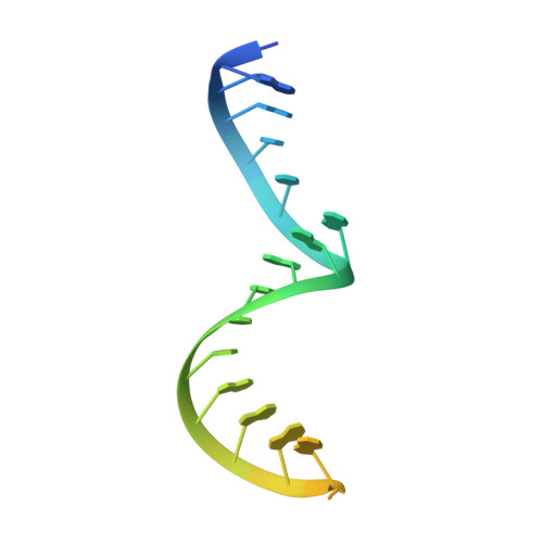
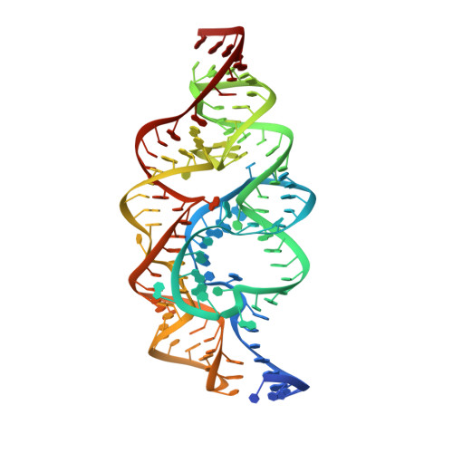
An in vitro evolved glmS ribozyme has the wild-type fold but loses coenzyme dependence.

Lau, M.W.Ferre-D'Amare, A.R.

(2013) Nat Chem Biol 9: 805-810

  • DOI: https://doi.org/10.1038/nchembio.1360
  • Primary Citation of Related Structures:  
    4MEG, 4MEH

  • PubMed Abstract: 

    Uniquely among known ribozymes, the glmS ribozyme-riboswitch requires a small-molecule coenzyme, glucosamine-6-phosphate (GlcN6P). Although consistent with its gene-regulatory function, the use of GlcN6P is unexpected because all of the other characterized self-cleaving ribozymes use RNA functional groups or divalent cations for catalysis. To determine what active site features make this ribozyme reliant on GlcN6P and to evaluate whether it might have evolved from a coenzyme-independent ancestor, we isolated a GlcN6P-independent variant through in vitro selection. Three active site mutations suffice to generate a highly reactive RNA that adopts the wild-type fold but uses divalent cations for catalysis and is insensitive to GlcN6P. Biochemical and crystallographic comparisons of wild-type and mutant ribozymes show that a handful of functional groups fine-tune the RNA to be either coenzyme or cation dependent. These results indicate that a few mutations can confer new biochemical activities on structured RNAs. Thus, families of structurally related ribozymes with divergent function may exist.


  • Organizational Affiliation

    National Heart, Lung and Blood Institute, Bethesda, Maryland, USA.


Macromolecules
Find similar nucleic acids by:  (by identity cutoff)  |  3D Structure
Entity ID: 1
MoleculeChains LengthOrganismImage
glmS triple mutant ribozyme27N/A
Sequence Annotations
Expand
  • Reference Sequence
Find similar nucleic acids by:  (by identity cutoff)  |  3D Structure
Entity ID: 2
MoleculeChains LengthOrganismImage
(121-MER) ribozyme126N/A
Sequence Annotations
Expand
  • Reference Sequence
Small Molecules
Ligands 1 Unique
IDChains Name / Formula / InChI Key2D Diagram3D Interactions
MG
Query on MG

Download Ideal Coordinates CCD File 
C [auth A]
D [auth B]
E [auth B]
F [auth B]
G [auth B]
C [auth A],
D [auth B],
E [auth B],
F [auth B],
G [auth B],
H [auth B],
I [auth B],
J [auth B],
K [auth B],
L [auth B],
M [auth B],
N [auth B],
O [auth B],
P [auth B],
Q [auth B],
R [auth B]
MAGNESIUM ION
Mg
JLVVSXFLKOJNIY-UHFFFAOYSA-N
Experimental Data & Validation

Experimental Data

  • Method: X-RAY DIFFRACTION
  • Resolution: 3.10 Å
  • R-Value Free: 0.252 
  • R-Value Work: 0.197 
  • R-Value Observed: 0.197 
  • Space Group: P 21 21 2
Unit Cell:
Length ( Å )Angle ( ˚ )
a = 166.9α = 90
b = 40.4β = 90
c = 73.6γ = 90
Software Package:
Software NamePurpose
HKL-2000data collection
PHASERphasing
CNSrefinement
HKL-2000data reduction
HKL-2000data scaling

Structure Validation

View Full Validation Report


Entry History 

Deposition Data

Revision History  (Full details and data files)

  • Version 1.0: 2013-10-16
    Type: Initial release
  • Version 1.1: 2013-11-27
    Changes: Database references
  • Version 1.2: 2023-09-20
    Changes: Data collection, Database references, Derived calculations, Refinement description