4M54

The structure of the staphyloferrin B precursor biosynthetic enzyme SbnB bound to N-(1-amino-1-carboxyl-2-ethyl)-glutamic acid and NADH


Experimental Data Snapshot

  • Method: X-RAY DIFFRACTION
  • Resolution: 2.36 Å
  • R-Value Free: 0.258 
  • R-Value Work: 0.223 
  • R-Value Observed: 0.227 

wwPDB Validation   3D Report Full Report


Ligand Structure Quality Assessment 


This is version 1.2 of the entry. See complete history


Literature

Synthesis of L-2,3-diaminopropionic acid, a siderophore and antibiotic precursor.

Kobylarz, M.J.Grigg, J.C.Takayama, S.J.Rai, D.K.Heinrichs, D.E.Murphy, M.E.

(2014) Chem Biol 21: 379-388

  • DOI: https://doi.org/10.1016/j.chembiol.2013.12.011
  • Primary Citation of Related Structures:  
    4M54, 4MP3, 4MP6, 4MP8, 4MPD

  • PubMed Abstract: 

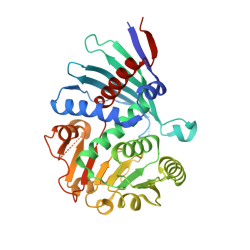
    L-2,3-diaminopropionic acid (L-Dap) is an amino acid that is a precursor of antibiotics and staphyloferrin B a siderophore produced by Staphylococcus aureus. SbnA and SbnB are encoded by the staphyloferrin B biosynthetic gene cluster and are implicated in L-Dap biosynthesis. We demonstrate here that SbnA uses PLP and substrates O-phospho-L-serine and L-glutamate to produce a metabolite N-(1-amino-1-carboxyl-2-ethyl)-glutamic acid (ACEGA). SbnB is shown to use NAD(+) to oxidatively hydrolyze ACEGA to yield α-ketoglutarate and L-Dap. Also, we describe crystal structures of SbnB in complex with NADH and ACEGA as well as with NAD(+) and α-ketoglutarate to reveal the residues required for substrate binding, oxidation, and hydrolysis. SbnA and SbnB contribute to the iron sparing response of S. aureus that enables staphyloferrin B biosynthesis in the absence of an active tricarboxylic acid cycle.


  • Organizational Affiliation

    Department of Microbiology and Immunology, Life Sciences Institute, The University of British Columbia, Vancouver, BC V6T 1Z3, Canada.


Macromolecules
Find similar proteins by:  (by identity cutoff)  |  3D Structure
Entity ID: 1
MoleculeChains Sequence LengthOrganismDetailsImage
Putative ornithine cyclodeaminase339Staphylococcus aureus subsp. aureus MW2Mutation(s): 0 
Gene Names: sbnB
UniProt
Find proteins for A0A0H3K9Y6 (Staphylococcus aureus (strain Newman))
Explore A0A0H3K9Y6 
Go to UniProtKB:  A0A0H3K9Y6
Entity Groups  
Sequence Clusters30% Identity50% Identity70% Identity90% Identity95% Identity100% Identity
UniProt GroupA0A0H3K9Y6
Sequence Annotations
Expand
  • Reference Sequence
Small Molecules
Ligands 3 Unique
IDChains Name / Formula / InChI Key2D Diagram3D Interactions
NAI
Query on NAI

Download Ideal Coordinates CCD File 
B [auth A]1,4-DIHYDRONICOTINAMIDE ADENINE DINUCLEOTIDE
C21 H29 N7 O14 P2
BOPGDPNILDQYTO-NNYOXOHSSA-N
AE5
Query on AE5

Download Ideal Coordinates CCD File 
D [auth A]N-[(2S)-2-amino-2-carboxyethyl]-L-glutamic acid
C8 H14 N2 O6
XYQHCOGLGSNTNV-WHFBIAKZSA-N
CL
Query on CL

Download Ideal Coordinates CCD File 
C [auth A]CHLORIDE ION
Cl
VEXZGXHMUGYJMC-UHFFFAOYSA-M
Experimental Data & Validation

Experimental Data

  • Method: X-RAY DIFFRACTION
  • Resolution: 2.36 Å
  • R-Value Free: 0.258 
  • R-Value Work: 0.223 
  • R-Value Observed: 0.227 
  • Space Group: P 41 21 2
Unit Cell:
Length ( Å )Angle ( ˚ )
a = 63.116α = 90
b = 63.116β = 90
c = 159.413γ = 90
Software Package:
Software NamePurpose
DENZOdata reduction
SCALEPACKdata scaling
PHENIXrefinement
PDB_EXTRACTdata extraction
HKL-3000data collection
HKL-3000data reduction
HKL-3000data scaling
PHENIXphasing

Structure Validation

View Full Validation Report


Ligand Structure Quality Assessment 


Entry History 

Deposition Data

Revision History  (Full details and data files)

  • Version 1.0: 2014-07-09
    Type: Initial release
  • Version 1.1: 2017-11-15
    Changes: Refinement description
  • Version 1.2: 2024-02-28
    Changes: Data collection, Database references, Derived calculations