4LWM

Crystal structure of methionine sulfoxide reductase U16C/E55D from clostridium oremlandii with methionie sulfoxide


Experimental Data Snapshot

  • Method: X-RAY DIFFRACTION
  • Resolution: 1.80 Å
  • R-Value Free: 0.223 
  • R-Value Work: 0.179 
  • R-Value Observed: 0.181 

wwPDB Validation   3D Report Full Report


Ligand Structure Quality Assessment 


This is version 2.1 of the entry. See complete history


Literature

Structural analysis of 1-Cys type selenoprotein methionine sulfoxide reductase A

Lee, E.H.Kwak, G.H.Kim, M.J.Kim, H.Y.Hwang, K.Y.

(2014) Arch Biochem Biophys 545: 1-8

  • DOI: https://doi.org/10.1016/j.abb.2013.12.024
  • Primary Citation of Related Structures:  
    4LWJ, 4LWK, 4LWL, 4LWM

  • PubMed Abstract: 

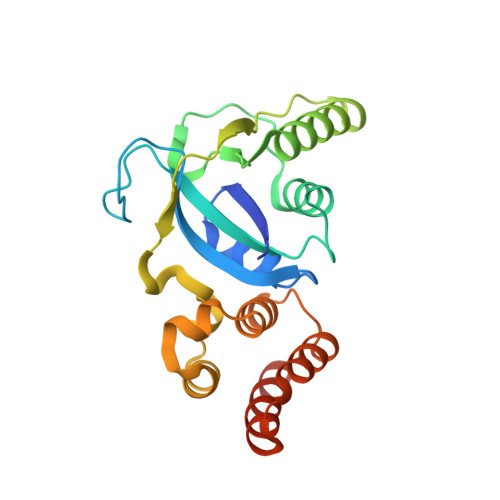
    Methionine sulfoxide reductase A (MsrA) reduces free and protein-based methionine-S-sulfoxide to methionine. Structures of 1-Cys MsrAs lacking a resolving Cys, which interacts with catalytic Cys, are unknown. In addition, no structural information on selenocysteine (Sec)-containing MsrA enzymes has been reported. In this work, we determined the crystal structures of 1-Cys type selenoprotein MsrA from Clostridium oremlandii at 1.6-1.8Å, including the reduced, oxidized (sulfenic acid), and substrate-bound forms. The overall structure of Clostridium MsrA, consisting of ten α-helices and six β-strands, folds into a catalytic domain and a novel helical domain absent from other known MsrA structures. The helical domain, containing five helices, tightly interacts with the catalytic domain, and is likely critical for catalytic activity due to its association with organizing the active site. This helical domain is also conserved in several selenoprotein MsrAs. Our structural analysis reveals that the side chain length of Glu55 is critical for the proton donor function of this residue. Our structures also provide insights into the architecture of the 1-Cys MsrA active site and the roles of active site residues in substrate recognition and catalysis.


  • Organizational Affiliation

    Division of Biotechnology, College of Life Sciences and Biotechnology, Korea University, Seoul 136-701, Republic of Korea.


Macromolecules
Find similar proteins by:  (by identity cutoff)  |  3D Structure
Entity ID: 1
MoleculeChains Sequence LengthOrganismDetailsImage
Peptide methionine sulfoxide reductase MsrA217Alkaliphilus oremlandii OhILAsMutation(s): 2 
Gene Names: msrA
EC: 1.8.4.11
UniProt
Find proteins for A8MI53 (Alkaliphilus oremlandii (strain OhILAs))
Explore A8MI53 
Go to UniProtKB:  A8MI53
Entity Groups  
Sequence Clusters30% Identity50% Identity70% Identity90% Identity95% Identity100% Identity
UniProt GroupA8MI53
Sequence Annotations
Expand
  • Reference Sequence
Small Molecules
Experimental Data & Validation

Experimental Data

  • Method: X-RAY DIFFRACTION
  • Resolution: 1.80 Å
  • R-Value Free: 0.223 
  • R-Value Work: 0.179 
  • R-Value Observed: 0.181 
  • Space Group: P 32 2 1
Unit Cell:
Length ( Å )Angle ( ˚ )
a = 80.849α = 90
b = 80.849β = 90
c = 66.921γ = 120
Software Package:
Software NamePurpose
MOLREPphasing
PHENIXrefinement
DENZOdata reduction
SCALEPACKdata scaling
HKL-2000data scaling

Structure Validation

View Full Validation Report


Ligand Structure Quality Assessment 


Entry History 

Deposition Data

Revision History  (Full details and data files)

  • Version 1.0: 2014-06-11
    Type: Initial release
  • Version 1.1: 2017-11-15
    Changes: Advisory, Refinement description
  • Version 2.0: 2019-08-07
    Changes: Data collection, Database references, Non-polymer description, Structure summary
  • Version 2.1: 2023-11-08
    Changes: Advisory, Data collection, Database references, Derived calculations, Refinement description