4KCW

Pyruvate kinase (PYK) from Trypanosoma brucei soaked with oxalate


Experimental Data Snapshot

  • Method: X-RAY DIFFRACTION
  • Resolution: 2.50 Å
  • R-Value Free: 0.202 
  • R-Value Work: 0.160 
  • R-Value Observed: 0.162 

wwPDB Validation   3D Report Full Report


Ligand Structure Quality Assessment 


This is version 1.3 of the entry. See complete history


Literature

Pyruvate kinases have an intrinsic and conserved decarboxylase activity.

Zhong, W.Morgan, H.P.Nowicki, M.W.McNae, I.W.Yuan, M.Bella, J.Michels, P.A.Fothergill-Gilmore, L.A.Walkinshaw, M.D.

(2014) Biochem J 458: 301-311

  • DOI: https://doi.org/10.1042/BJ20130790
  • Primary Citation of Related Structures:  
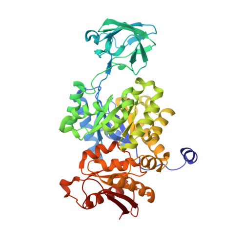
    4KCT, 4KCU, 4KCV, 4KCW

  • PubMed Abstract: 

    The phosphotransfer mechanism of PYKs (pyruvate kinases) has been studied in detail, but the mechanism of the intrinsic decarboxylase reaction catalysed by PYKs is still unknown. 1H NMR was used in the present study to follow OAA (oxaloacetate) decarboxylation by trypanosomatid and human PYKs confirming that the decarboxylase activity is conserved across distantly related species. Crystal structures of TbPYK (Trypanosoma brucei PYK) complexed with the product of the decarboxylase reaction (pyruvate), and a series of substrate analogues (D-malate, 2-oxoglutarate and oxalate) show that the OAA analogues bind to the kinase active site with similar binding modes, confirming that both decarboxylase and kinase activities share a common site for substrate binding and catalysis. Decarboxylation of OAA as monitored by NMR for TbPYK has a relatively low turnover with values of 0.86 s-1 and 1.47 s-1 in the absence and presence of F26BP (fructose 2,6-bisphosphate) respectively. Human M1PYK (M1 isoform of PYK) has a measured turnover value of 0.50 s-1. The X-ray structures explain why the decarboxylation activity is specific for OAA and is not general for α-oxo acid analogues. Conservation of the decarboxylase reaction across divergent species is a consequence of piggybacking on the conserved kinase mechanism which requires a stabilized enol intermediate.


  • Organizational Affiliation

    *Centre for Translational and Chemical Biology, School of Biological Sciences, University of Edinburgh, Michael Swann Building, The King's Buildings, Mayfield Road, Edinburgh EH9 3JR, U.K.


Macromolecules
Find similar proteins by:  (by identity cutoff)  |  3D Structure
Entity ID: 1
MoleculeChains Sequence LengthOrganismDetailsImage
Pyruvate kinase 1
A, B
499Trypanosoma brucei bruceiMutation(s): 0 
Gene Names: PYK1
EC: 2.7.1.40
UniProt
Find proteins for P30615 (Trypanosoma brucei brucei)
Explore P30615 
Go to UniProtKB:  P30615
Entity Groups  
Sequence Clusters30% Identity50% Identity70% Identity90% Identity95% Identity100% Identity
UniProt GroupP30615
Sequence Annotations
Expand
  • Reference Sequence
Small Molecules
Ligands 5 Unique
IDChains Name / Formula / InChI Key2D Diagram3D Interactions
FDP
Query on FDP

Download Ideal Coordinates CCD File 
F [auth A],
J [auth B]
2,6-di-O-phosphono-beta-D-fructofuranose
C6 H14 O12 P2
YXWOAJXNVLXPMU-ZXXMMSQZSA-N
GOL
Query on GOL

Download Ideal Coordinates CCD File 
K [auth B],
L [auth B],
M [auth B]
GLYCEROL
C3 H8 O3
PEDCQBHIVMGVHV-UHFFFAOYSA-N
OXL
Query on OXL

Download Ideal Coordinates CCD File 
E [auth A],
I [auth B]
OXALATE ION
C2 O4
MUBZPKHOEPUJKR-UHFFFAOYSA-L
K
Query on K

Download Ideal Coordinates CCD File 
D [auth A],
H [auth B]
POTASSIUM ION
K
NPYPAHLBTDXSSS-UHFFFAOYSA-N
MG
Query on MG

Download Ideal Coordinates CCD File 
C [auth A],
G [auth B]
MAGNESIUM ION
Mg
JLVVSXFLKOJNIY-UHFFFAOYSA-N
Experimental Data & Validation

Experimental Data

  • Method: X-RAY DIFFRACTION
  • Resolution: 2.50 Å
  • R-Value Free: 0.202 
  • R-Value Work: 0.160 
  • R-Value Observed: 0.162 
  • Space Group: I 2 2 2
Unit Cell:
Length ( Å )Angle ( ˚ )
a = 103.88α = 90
b = 108.56β = 90
c = 264.53γ = 90
Software Package:
Software NamePurpose
DNAdata collection
PHASERphasing
REFMACrefinement
MOSFLMdata reduction
SCALAdata scaling

Structure Validation

View Full Validation Report


Ligand Structure Quality Assessment 


Entry History 

Deposition Data

Revision History  (Full details and data files)

  • Version 1.0: 2014-01-08
    Type: Initial release
  • Version 1.1: 2014-02-26
    Changes: Database references
  • Version 1.2: 2020-07-29
    Type: Remediation
    Reason: Carbohydrate remediation
    Changes: Data collection, Derived calculations, Structure summary
  • Version 1.3: 2024-02-28
    Changes: Data collection, Database references, Structure summary