4KBB

Structure of Botulinum neurotoxin B binding domain in complex with both synaptotagmin II and GD1a


Experimental Data Snapshot

  • Method: X-RAY DIFFRACTION
  • Resolution: 2.30 Å
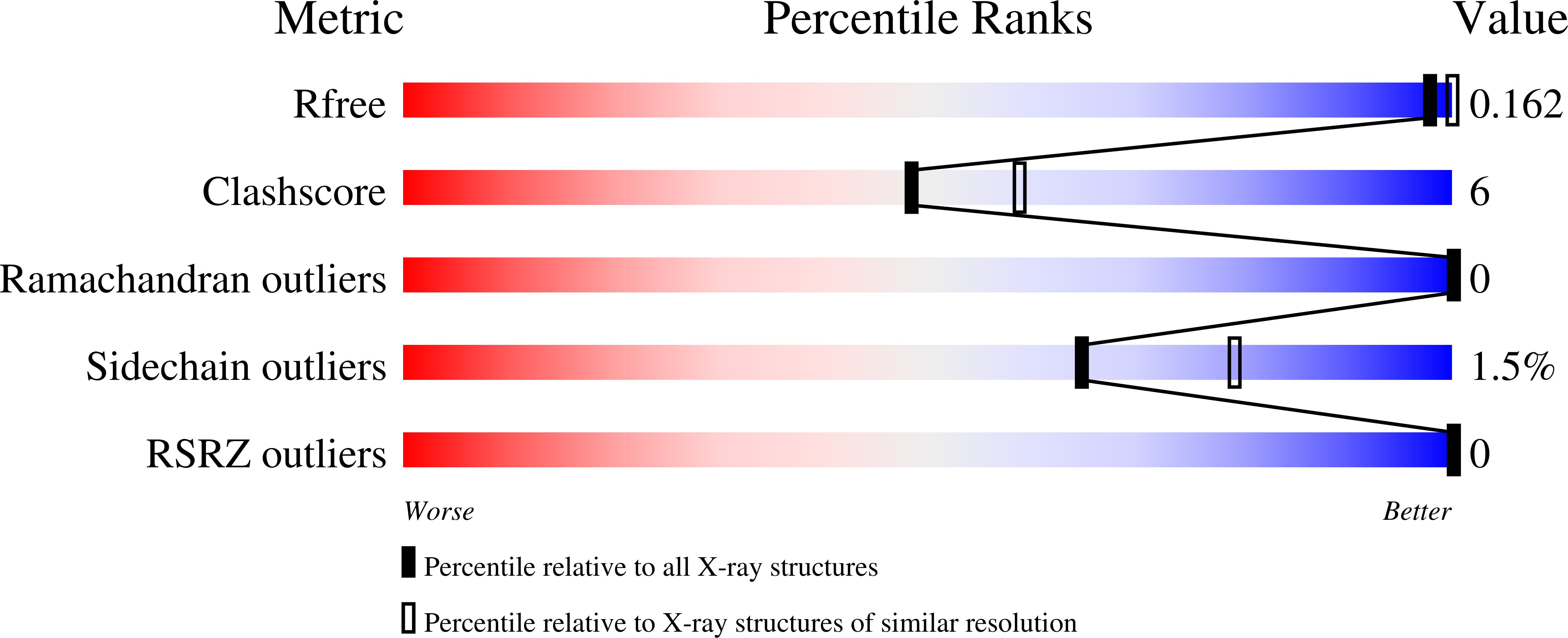
  • R-Value Free: 0.189 
  • R-Value Work: 0.156 
  • R-Value Observed: 0.159 

wwPDB Validation   3D Report Full Report


This is version 2.1 of the entry. See complete history


Literature

Structure of dual receptor binding to botulinum neurotoxin B.

Berntsson, R.P.Peng, L.Dong, M.Stenmark, P.

(2013) Nat Commun 4: 2058-2058

  • DOI: https://doi.org/10.1038/ncomms3058
  • Primary Citation of Related Structures:  
    4KBB

  • PubMed Abstract: 

    Botulinum neurotoxins are highly toxic, and bind two receptors to achieve their high affinity and specificity for neurons. Here we present the first structure of a botulinum neurotoxin bound to both its receptors. We determine the 2.3-Å structure of a ternary complex of botulinum neurotoxin type B bound to both its protein receptor synaptotagmin II and its ganglioside receptor GD1a. We show that there is no direct contact between the two receptors, and that the binding affinity towards synaptotagmin II is not influenced by the presence of GD1a. The interactions of botulinum neurotoxin type B with the sialic acid 5 moiety of GD1a are important for the ganglioside selectivity. The structure demonstrates that the protein receptor and the ganglioside receptor occupy nearby but separate binding sites, thus providing two independent anchoring points.


  • Organizational Affiliation

    Department of Biochemistry and Biophysics, Stockholm University, Stockholm 10691, Sweden.


Macromolecules
Find similar proteins by:  (by identity cutoff)  |  3D Structure
Entity ID: 1
MoleculeChains Sequence LengthOrganismDetailsImage
Botulinum neurotoxin type B
A, B
459Clostridium botulinumMutation(s): 0 
Gene Names: botB
EC: 3.4.24.69
UniProt
Find proteins for P10844 (Clostridium botulinum)
Explore P10844 
Go to UniProtKB:  P10844
Entity Groups  
Sequence Clusters30% Identity50% Identity70% Identity90% Identity95% Identity100% Identity
UniProt GroupP10844
Sequence Annotations
Expand
  • Reference Sequence
Find similar proteins by:  (by identity cutoff)  |  3D Structure
Entity ID: 2
MoleculeChains Sequence LengthOrganismDetailsImage
Synaptotagmin-2
C, D
68Mus musculusMutation(s): 0 
Gene Names: Syt2
UniProt
Find proteins for P46097 (Mus musculus)
Explore P46097 
Go to UniProtKB:  P46097
Entity Groups  
Sequence Clusters30% Identity50% Identity70% Identity90% Identity95% Identity100% Identity
UniProt GroupP46097
Sequence Annotations
Expand
  • Reference Sequence
Oligosaccharides

Help

Entity ID: 3
MoleculeChains Length2D Diagram Glycosylation3D Interactions
N-acetyl-alpha-neuraminic acid-(2-3)-beta-D-galactopyranose-(1-3)-2-acetamido-2-deoxy-beta-D-galactopyranose-(1-4)-[N-acetyl-alpha-neuraminic acid-(2-3)]beta-D-galactopyranose-(1-4)-beta-D-glucopyranose
E, F
6N/A
Glycosylation Resources
GlyTouCan:  G46677TE
GlyCosmos:  G46677TE
GlyGen:  G46677TE
Experimental Data & Validation

Experimental Data

  • Method: X-RAY DIFFRACTION
  • Resolution: 2.30 Å
  • R-Value Free: 0.189 
  • R-Value Work: 0.156 
  • R-Value Observed: 0.159 
  • Space Group: P 1 21 1
Unit Cell:
Length ( Å )Angle ( ˚ )
a = 47.163α = 90
b = 158.237β = 108.44
c = 75.244γ = 90
Software Package:
Software NamePurpose
SCALAdata scaling
PHENIXrefinement
PDB_EXTRACTdata extraction
XSCALEdata scaling

Structure Validation

View Full Validation Report


Entry History 

Deposition Data

Revision History  (Full details and data files)

  • Version 1.0: 2013-07-03
    Type: Initial release
  • Version 1.1: 2013-07-24
    Changes: Database references
  • Version 2.0: 2020-07-29
    Type: Remediation
    Reason: Carbohydrate remediation
    Changes: Advisory, Atomic model, Data collection, Database references, Derived calculations, Structure summary
  • Version 2.1: 2024-02-28
    Changes: Advisory, Data collection, Database references, Structure summary