4K9F

Neutron structure of Perdeuterated Rubredoxin refined against 1.75 resolution data collected on the new IMAGINE instrument at HFIR, ORNL


Experimental Data Snapshot

  • Method: NEUTRON DIFFRACTION
  • Resolution: 1.75 Å
  • R-Value Free: 0.241 
  • R-Value Work: 0.199 
  • R-Value Observed: 0.203 

wwPDB Validation   3D Report Full Report


This is version 1.2 of the entry. See complete history


Literature

The IMAGINE instrument: first neutron protein structure and new capabilities for neutron macromolecular crystallography.

Meilleur, F.Munshi, P.Robertson, L.Stoica, A.D.Crow, L.Kovalevsky, A.Koritsanszky, T.Chakoumakos, B.C.Blessing, R.Myles, D.A.

(2013) Acta Crystallogr D Biol Crystallogr 69: 2157-2160

  • DOI: https://doi.org/10.1107/S0907444913019604
  • Primary Citation of Related Structures:  
    4K9F

  • PubMed Abstract: 

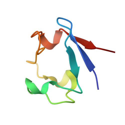
    The first high-resolution neutron protein structure of perdeuterated rubredoxin from Pyrococcus furiosus (PfRd) determined using the new IMAGINE macromolecular neutron crystallography instrument at the Oak Ridge National Laboratory is reported. Neutron diffraction data extending to 1.65 Å resolution were collected from a relatively small 0.7 mm(3) PfRd crystal using 2.5 d (60 h) of beam time. The refined structure contains 371 out of 391, or 95%, of the D atoms of the protein and 58 solvent molecules. The IMAGINE instrument is designed to provide neutron data at or near atomic resolution (1.5 Å) from crystals with volume <1.0 mm(3) and with unit-cell edges <100 Å. Beamline features include novel elliptical focusing mirrors that deliver neutrons into a 2.0 × 3.2 mm focal spot at the sample position with full-width vertical and horizontal divergences of 0.5 and 0.6°, respectively. Variable short- and long-wavelength cutoff optics provide automated exchange between multiple-wavelength configurations (λmin = 2.0, 2.8, 3.3 Å to λmax = 3.0, 4.0, 4.5, ∼20 Å). These optics produce a more than 20-fold increase in the flux density at the sample and should help to enable more routine collection of high-resolution data from submillimetre-cubed crystals. Notably, the crystal used to collect these PfRd data was 5-10 times smaller than those previously reported.


  • Organizational Affiliation

    Department of Molecular and Structural Biochemistry, NCSU, Raleigh, NC 27695, USA.


Macromolecules
Find similar proteins by:  (by identity cutoff)  |  3D Structure
Entity ID: 1
MoleculeChains Sequence LengthOrganismDetailsImage
Rubredoxin54Pyrococcus furiosus DSM 3638Mutation(s): 0 
Gene Names: PF1282rub
UniProt
Find proteins for P24297 (Pyrococcus furiosus (strain ATCC 43587 / DSM 3638 / JCM 8422 / Vc1))
Explore P24297 
Go to UniProtKB:  P24297
Entity Groups  
Sequence Clusters30% Identity50% Identity70% Identity90% Identity95% Identity100% Identity
UniProt GroupP24297
Sequence Annotations
Expand
  • Reference Sequence
Small Molecules
Ligands 1 Unique
IDChains Name / Formula / InChI Key2D Diagram3D Interactions
FE
Query on FE

Download Ideal Coordinates CCD File 
B [auth A]FE (III) ION
Fe
VTLYFUHAOXGGBS-UHFFFAOYSA-N
Experimental Data & Validation

Experimental Data

  • Method: NEUTRON DIFFRACTION
  • Resolution: 1.75 Å
  • R-Value Free: 0.241 
  • R-Value Work: 0.199 
  • R-Value Observed: 0.203 
  • Space Group: P 21 21 21
Unit Cell:
Length ( Å )Angle ( ˚ )
a = 33.822α = 90
b = 34.803β = 90
c = 43.351γ = 90
Software Package:
Software NamePurpose
Quasi-Lauedata collection
PHENIXrefinement
LAUEGENdata reduction
SCALAdata scaling

Structure Validation

View Full Validation Report


Entry History 

Deposition Data

Revision History  (Full details and data files)

  • Version 1.0: 2013-12-04
    Type: Initial release
  • Version 1.1: 2018-06-13
    Changes: Data collection
  • Version 1.2: 2023-09-20
    Changes: Data collection, Database references, Derived calculations, Refinement description