4JIP

Crystal structure of glycerol trinitrate reductase NerA from Agrobacterium radiobacter in complex with 4-hydroxybenzaldehyde


Experimental Data Snapshot

  • Method: X-RAY DIFFRACTION
  • Resolution: 2.29 Å
  • R-Value Free: 0.239 
  • R-Value Work: 0.194 
  • R-Value Observed: 0.197 

wwPDB Validation   3D Report Full Report


Ligand Structure Quality Assessment 


This is version 1.2 of the entry. See complete history


Literature

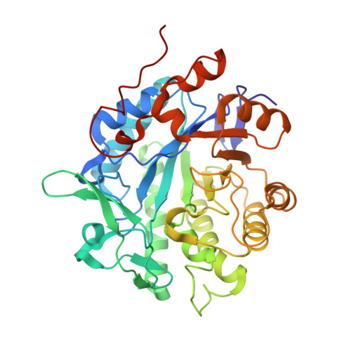
The Structure of Glycerol Trinitrate Reductase NerA from Agrobacterium radiobacter Reveals the Molecular Reason for Nitro- and Ene-Reductase Activity in OYE Homologues.

Oberdorfer, G.Binter, A.Wallner, S.Durchschein, K.Hall, M.Faber, K.Macheroux, P.Gruber, K.

(2013) Chembiochem 14: 836-845

  • DOI: https://doi.org/10.1002/cbic.201300136
  • Primary Citation of Related Structures:  
    4JIC, 4JIP, 4JIQ

  • PubMed Abstract: 

    In recent years, Old Yellow Enzymes (OYEs) and their homologues have found broad application in the efficient asymmetric hydrogenation of activated C=C bonds with high selectivities and yields. Members of this class of enzymes have been found in many different organisms and are rather diverse on the sequence level, with pairwise identities as low as 20 %, but they exhibit significant structural similarities with the adoption of a conserved (αβ)(8)-barrel fold. Some OYEs have been shown not only to reduce C=C double bonds, but also to be capable of reducing nitro groups in both saturated and unsaturated substrates. In order to understand this dual activity we determined and analyzed X-ray crystal structures of NerA from Agrobacterium radiobacter, both in its apo form and in complex with 4-hydroxybenzaldehyde and with 1-nitro-2-phenylpropene. These structures, together with spectroscopic studies of substrate binding to several OYEs, indicate that nitro-containing substrates can bind to OYEs in different binding modes, one of which leads to C=C double bond reduction and the other to nitro group reduction.


  • Organizational Affiliation

    ACIB--Austrian Centre of Industrial Biotechnology, Petergasse 14, 8010 Graz, Austria.


Macromolecules
Find similar proteins by:  (by identity cutoff)  |  3D Structure
Entity ID: 1
MoleculeChains Sequence LengthOrganismDetailsImage
GTN Reductase377Agrobacterium tumefaciensMutation(s): 0 
Gene Names: nerA
UniProt
Find proteins for O31246 (Rhizobium radiobacter)
Explore O31246 
Go to UniProtKB:  O31246
Entity Groups  
Sequence Clusters30% Identity50% Identity70% Identity90% Identity95% Identity100% Identity
UniProt GroupO31246
Sequence Annotations
Expand
  • Reference Sequence
Small Molecules
Ligands 2 Unique
IDChains Name / Formula / InChI Key2D Diagram3D Interactions
FMN
Query on FMN

Download Ideal Coordinates CCD File 
B [auth A]FLAVIN MONONUCLEOTIDE
C17 H21 N4 O9 P
FVTCRASFADXXNN-SCRDCRAPSA-N
HBA
Query on HBA

Download Ideal Coordinates CCD File 
C [auth A]P-HYDROXYBENZALDEHYDE
C7 H6 O2
RGHHSNMVTDWUBI-UHFFFAOYSA-N
Experimental Data & Validation

Experimental Data

  • Method: X-RAY DIFFRACTION
  • Resolution: 2.29 Å
  • R-Value Free: 0.239 
  • R-Value Work: 0.194 
  • R-Value Observed: 0.197 
  • Space Group: P 21 21 21
Unit Cell:
Length ( Å )Angle ( ˚ )
a = 58.91α = 90
b = 68.28β = 90
c = 90.06γ = 90
Software Package:
Software NamePurpose
MAR345dtbdata collection
PHASERphasing
PHENIXrefinement
MOSFLMdata reduction
SCALAdata scaling

Structure Validation

View Full Validation Report


Ligand Structure Quality Assessment 


Entry History 

Deposition Data

Revision History  (Full details and data files)

  • Version 1.0: 2013-05-08
    Type: Initial release
  • Version 1.1: 2013-05-15
    Changes: Database references
  • Version 1.2: 2023-09-20
    Changes: Data collection, Database references, Derived calculations, Refinement description