4JHP

The crystal structure of the RPGR RCC1-like domain in complex with PDE6D


Experimental Data Snapshot

  • Method: X-RAY DIFFRACTION
  • Resolution: 1.90 Å
  • R-Value Free: 0.221 
  • R-Value Work: 0.200 
  • R-Value Observed: 0.201 

wwPDB Validation   3D Report Full Report


This is version 1.2 of the entry. See complete history


Literature

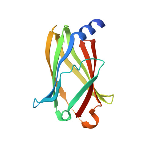
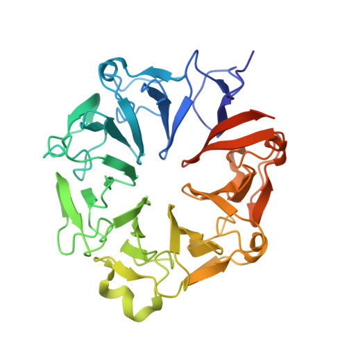
The interplay between RPGR, PDE-delta and Arl2/3 regulate the ciliary targeting of farnesylated cargo.

Watzlich, D.Vetter, I.Gotthardt, K.Miertzschke, M.Chen, Y.X.Wittinghofer, A.Ismail, S.

(2013) EMBO Rep 14: 465-472

  • DOI: https://doi.org/10.1038/embor.2013.37
  • Primary Citation of Related Structures:  
    4JHN, 4JHP

  • PubMed Abstract: 

    Defects in primary cilia result in human diseases known as ciliopathies. The retinitis pigmentosa GTPase regulator (RPGR), mutated in the most severe form of the eye disease, is located at the transition zone of the ciliary organelle. The RPGR-interacting partner PDEδ is involved in trafficking of farnesylated ciliary cargo, but the significance of this interaction is unknown. The crystal structure of the propeller domain of RPGR shows the location of patient mutations and how they perturb the structure. The RPGR·PDEδ complex structure shows PDEδ on a highly conserved surface patch of RPGR. Biochemical experiments and structural considerations show that RPGR can bind with high affinity to cargo-loaded PDEδ and exposes the Arl2/Arl3-binding site on PDEδ. On the basis of these results, we propose a model where RPGR is acting as a scaffold protein recruiting cargo-loaded PDEδ and Arl3 to release lipidated cargo into cilia.


  • Organizational Affiliation

    Structural Biology Group, Max Planck Institute for Molecular Physiology, Otto-Hahn-Strasse 11, Dortmund 44227, Germany.


Macromolecules
Find similar proteins by:  (by identity cutoff)  |  3D Structure
Entity ID: 1
MoleculeChains Sequence LengthOrganismDetailsImage
Retinal rod rhodopsin-sensitive cGMP 3',5'-cyclic phosphodiesterase subunit deltaA [auth B]152Homo sapiensMutation(s): 0 
Gene Names: PDE6DPDED
UniProt & NIH Common Fund Data Resources
Find proteins for O43924 (Homo sapiens)
Explore O43924 
Go to UniProtKB:  O43924
PHAROS:  O43924
GTEx:  ENSG00000156973 
Entity Groups  
Sequence Clusters30% Identity50% Identity70% Identity90% Identity95% Identity100% Identity
UniProt GroupO43924
Sequence Annotations
Expand
  • Reference Sequence
Find similar proteins by:  (by identity cutoff)  |  3D Structure
Entity ID: 2
MoleculeChains Sequence LengthOrganismDetailsImage
X-linked retinitis pigmentosa GTPase regulatorB [auth C]391Homo sapiensMutation(s): 0 
Gene Names: RPGRRP3XLRP3
UniProt & NIH Common Fund Data Resources
Find proteins for Q92834 (Homo sapiens)
Explore Q92834 
Go to UniProtKB:  Q92834
PHAROS:  Q92834
GTEx:  ENSG00000156313 
Entity Groups  
Sequence Clusters30% Identity50% Identity70% Identity90% Identity95% Identity100% Identity
UniProt GroupQ92834
Sequence Annotations
Expand
  • Reference Sequence
Experimental Data & Validation

Experimental Data

  • Method: X-RAY DIFFRACTION
  • Resolution: 1.90 Å
  • R-Value Free: 0.221 
  • R-Value Work: 0.200 
  • R-Value Observed: 0.201 
  • Space Group: C 1 2 1
Unit Cell:
Length ( Å )Angle ( ˚ )
a = 133.83α = 90
b = 58.78β = 123.32
c = 92.11γ = 90
Software Package:
Software NamePurpose
PROdata collection
MOLREPphasing
REFMACrefinement
XDSdata reduction
XSCALEdata scaling

Structure Validation

View Full Validation Report


Entry History 

Deposition Data

Revision History  (Full details and data files)

  • Version 1.0: 2013-04-17
    Type: Initial release
  • Version 1.1: 2013-06-19
    Changes: Database references
  • Version 1.2: 2023-09-20
    Changes: Data collection, Database references, Refinement description