4JBS

Crystal structure of the human Endoplasmic Reticulum Aminopeptidase 2 in complex with PHOSPHINIC PSEUDOTRIPEPTIDE inhibitor.


Experimental Data Snapshot

  • Method: X-RAY DIFFRACTION
  • Resolution: 2.79 Å
  • R-Value Free: 0.278 
  • R-Value Work: 0.206 
  • R-Value Observed: 0.210 

wwPDB Validation   3D Report Full Report


Ligand Structure Quality Assessment 


This is version 2.1 of the entry. See complete history


Literature

Rationally designed inhibitor targeting antigen-trimming aminopeptidases enhances antigen presentation and cytotoxic T-cell responses.

Zervoudi, E.Saridakis, E.Birtley, J.R.Seregin, S.S.Reeves, E.Kokkala, P.Aldhamen, Y.A.Amalfitano, A.Mavridis, I.M.James, E.Georgiadis, D.Stratikos, E.

(2013) Proc Natl Acad Sci U S A 110: 19890-19895

  • DOI: https://doi.org/10.1073/pnas.1309781110
  • Primary Citation of Related Structures:  
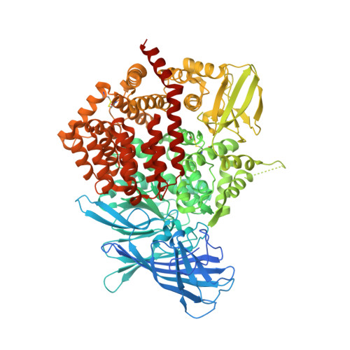
    4JBS

  • PubMed Abstract: 

    Intracellular aminopeptidases endoplasmic reticulum aminopeptidases 1 and 2 (ERAP1 and ERAP2), and as well as insulin-regulated aminopeptidase (IRAP) process antigenic epitope precursors for loading onto MHC class I molecules and regulate the adaptive immune response. Their activity greatly affects the antigenic peptide repertoire presented to cytotoxic T lymphocytes and as a result can regulate cytotoxic cellular responses contributing to autoimmunity or immune evasion by viruses and cancer cells. Therefore, pharmacological regulation of their activity is a promising avenue for modulating the adaptive immune response with possible applications in controlling autoimmunity, in boosting immune responses to pathogens, and in cancer immunotherapy. In this study we exploited recent structural and biochemical analysis of ERAP1 and ERAP2 to design and develop phosphinic pseudopeptide transition state analogs that can inhibit this family of enzymes with nM affinity. X-ray crystallographic analysis of one such inhibitor in complex with ERAP2 validated our design, revealing a canonical mode of binding in the active site of the enzyme, and highlighted the importance of the S2' pocket for achieving inhibitor potency. Antigen processing and presentation assays in HeLa and murine colon carcinoma (CT26) cells showed that these inhibitors induce increased cell-surface antigen presentation of transfected and endogenous antigens and enhance cytotoxic T-cell responses, indicating that these enzymes primarily destroy epitopes in those systems. This class of inhibitors constitutes a promising tool for controlling the cellular adaptive immune response in humans by modulating the antigen processing and presentation pathway.


  • Organizational Affiliation

    National Center for Scientific Research Demokritos, Agia Paraskevi 15310, Greece.


Macromolecules
Find similar proteins by:  (by identity cutoff)  |  3D Structure
Entity ID: 1
MoleculeChains Sequence LengthOrganismDetailsImage
Endoplasmic reticulum aminopeptidase 2
A, B
967Homo sapiensMutation(s): 0 
Gene Names: ERAP2LRAP
EC: 3.4.11
UniProt & NIH Common Fund Data Resources
Find proteins for Q6P179 (Homo sapiens)
Explore Q6P179 
Go to UniProtKB:  Q6P179
PHAROS:  Q6P179
GTEx:  ENSG00000164308 
Entity Groups  
Sequence Clusters30% Identity50% Identity70% Identity90% Identity95% Identity100% Identity
UniProt GroupQ6P179
Sequence Annotations
Expand
  • Reference Sequence
Oligosaccharides

Help

Entity ID: 2
MoleculeChains Length2D Diagram Glycosylation3D Interactions
2-acetamido-2-deoxy-beta-D-glucopyranose-(1-4)-2-acetamido-2-deoxy-beta-D-glucopyranose
C, D, F
2N-Glycosylation
Glycosylation Resources
GlyTouCan:  G42666HT
GlyCosmos:  G42666HT
GlyGen:  G42666HT
Entity ID: 3
MoleculeChains Length2D Diagram Glycosylation3D Interactions
alpha-D-mannopyranose-(1-4)-alpha-D-mannopyranose-(1-4)-2-acetamido-2-deoxy-beta-D-glucopyranose-(1-4)-2-acetamido-2-deoxy-beta-D-glucopyranose
E
4N-Glycosylation
Glycosylation Resources
GlyTouCan:  G81874LD
GlyCosmos:  G81874LD
GlyGen:  G81874LD
Small Molecules
Ligands 4 Unique
IDChains Name / Formula / InChI Key2D Diagram3D Interactions
P52
Query on P52

Download Ideal Coordinates CCD File 
K [auth A],
Q [auth B]
Nalpha-[(2S)-2-{[[(1R)-1-amino-3-phenylpropyl](hydroxy)phosphoryl]methyl}-4-methylpentanoyl]-L-tryptophanamide
C27 H37 N4 O4 P
QKFOTLXPIIESQI-IEZKXTBUSA-N
NAG
Query on NAG

Download Ideal Coordinates CCD File 
G [auth A],
H [auth A],
I [auth A],
N [auth B],
O [auth B]
2-acetamido-2-deoxy-beta-D-glucopyranose
C8 H15 N O6
OVRNDRQMDRJTHS-FMDGEEDCSA-N
IMD
Query on IMD

Download Ideal Coordinates CCD File 
L [auth A],
M [auth A]
IMIDAZOLE
C3 H5 N2
RAXXELZNTBOGNW-UHFFFAOYSA-O
ZN
Query on ZN

Download Ideal Coordinates CCD File 
J [auth A],
P [auth B]
ZINC ION
Zn
PTFCDOFLOPIGGS-UHFFFAOYSA-N
Binding Affinity Annotations 
IDSourceBinding Affinity
P52 PDBBind:  4JBS IC50: 11 (nM) from 1 assay(s)
BindingDB:  4JBS IC50: min: 11, max: 80 (nM) from 3 assay(s)
Experimental Data & Validation

Experimental Data

  • Method: X-RAY DIFFRACTION
  • Resolution: 2.79 Å
  • R-Value Free: 0.278 
  • R-Value Work: 0.206 
  • R-Value Observed: 0.210 
  • Space Group: P 1 21 1
Unit Cell:
Length ( Å )Angle ( ˚ )
a = 75.15α = 90
b = 134.77β = 90.28
c = 128.731γ = 90
Software Package:
Software NamePurpose
MOLREPphasing
PHENIXrefinement
MOSFLMdata reduction
SCALAdata scaling

Structure Validation

View Full Validation Report


Ligand Structure Quality Assessment 


Entry History 

Deposition Data

Revision History  (Full details and data files)

  • Version 1.0: 2013-12-11
    Type: Initial release
  • Version 1.1: 2013-12-25
    Changes: Database references
  • Version 1.2: 2017-11-15
    Changes: Advisory, Refinement description
  • Version 2.0: 2020-07-29
    Type: Remediation
    Reason: Carbohydrate remediation
    Changes: Advisory, Atomic model, Data collection, Database references, Derived calculations, Structure summary
  • Version 2.1: 2023-09-20
    Changes: Advisory, Data collection, Database references, Refinement description, Structure summary