4JAY

Crystal structure of P. aeruginosa MurB in complex with NADP+


Experimental Data Snapshot

  • Method: X-RAY DIFFRACTION
  • Resolution: 2.23 Å
  • R-Value Free: 0.258 
  • R-Value Work: 0.223 
  • R-Value Observed: 0.224 

wwPDB Validation   3D Report Full Report


Ligand Structure Quality Assessment 


This is version 1.1 of the entry. See complete history


Literature

Substrate Channel Flexibility in Pseudomonas aeruginosa MurB Accommodates Two Distinct Substrates.

Chen, M.W.Lohkamp, B.Schnell, R.Lescar, J.Schneider, G.

(2013) PLoS One 8: e66936-e66936

  • DOI: https://doi.org/10.1371/journal.pone.0066936
  • Primary Citation of Related Structures:  
    4JAY, 4JB1

  • PubMed Abstract: 

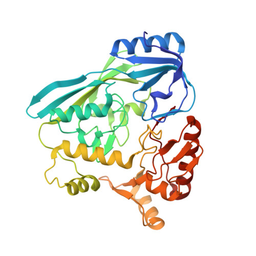
    Biosynthesis of UDP-N-acetylmuramic acid in bacteria is a committed step towards peptidoglycan production. In an NADPH- and FAD-dependent reaction, the UDP-N-acetylglucosamine-enolpyruvate reductase (MurB) reduces UDP-N-acetylglucosamine-enolpyruvate to UDP-N-acetylmuramic acid. We determined the three-dimensional structures of the ternary complex of Pseudomonas aeruginosa MurB with FAD and NADP(+) in two crystal forms to resolutions of 2.2 and 2.1 Å, respectively, to investigate the structural basis of the first half-reaction, hydride transfer from NADPH to FAD. The nicotinamide ring of NADP(+) stacks against the si face of the isoalloxazine ring of FAD, suggesting an unusual mode of hydride transfer to flavin. Comparison with the structure of the Escherichia coli MurB complex with UDP-N-acetylglucosamine-enolpyruvate shows that both substrates share the binding site located between two lobes of the substrate-binding domain III, consistent with a ping pong mechanism with sequential substrate binding. The nicotinamide and the enolpyruvyl moieties are strikingly well-aligned upon superimposition, both positioned for hydride transfer to and from FAD. However, flexibility of the substrate channel allows the non-reactive parts of the two substrates to bind in different conformations. A potassium ion in the active site may assist in substrate orientation and binding. These structural models should help in structure-aided drug design against MurB, which is essential for cell wall biogenesis and hence bacterial survival.


  • Organizational Affiliation

    Department of Medical Biochemistry and Biophysics, Karolinska Institutet, Stockholm, Sweden ; School of Biological Sciences, Nanyang Technological University, Singapore, Singapore.


Macromolecules
Find similar proteins by:  (by identity cutoff)  |  3D Structure
Entity ID: 1
MoleculeChains Sequence LengthOrganismDetailsImage
UDP-N-acetylenolpyruvoylglucosamine reductase
A, B, C, D
340Pseudomonas aeruginosa PAO1Mutation(s): 0 
Gene Names: murBPA2977
EC: 1.1.1.158
UniProt
Find proteins for Q9HZM7 (Pseudomonas aeruginosa (strain ATCC 15692 / DSM 22644 / CIP 104116 / JCM 14847 / LMG 12228 / 1C / PRS 101 / PAO1))
Explore Q9HZM7 
Go to UniProtKB:  Q9HZM7
Entity Groups  
Sequence Clusters30% Identity50% Identity70% Identity90% Identity95% Identity100% Identity
UniProt GroupQ9HZM7
Sequence Annotations
Expand
  • Reference Sequence
Small Molecules
Ligands 4 Unique
IDChains Name / Formula / InChI Key2D Diagram3D Interactions
FAD
Query on FAD

Download Ideal Coordinates CCD File 
E [auth A],
J [auth B],
N [auth C],
R [auth D]
FLAVIN-ADENINE DINUCLEOTIDE
C27 H33 N9 O15 P2
VWWQXMAJTJZDQX-UYBVJOGSSA-N
NAP
Query on NAP

Download Ideal Coordinates CCD File 
F [auth A],
K [auth B],
O [auth C],
S [auth D]
NADP NICOTINAMIDE-ADENINE-DINUCLEOTIDE PHOSPHATE
C21 H28 N7 O17 P3
XJLXINKUBYWONI-NNYOXOHSSA-N
B3P
Query on B3P

Download Ideal Coordinates CCD File 
G [auth A],
L [auth B],
P [auth C],
T [auth D]
2-[3-(2-HYDROXY-1,1-DIHYDROXYMETHYL-ETHYLAMINO)-PROPYLAMINO]-2-HYDROXYMETHYL-PROPANE-1,3-DIOL
C11 H26 N2 O6
HHKZCCWKTZRCCL-UHFFFAOYSA-N
K
Query on K

Download Ideal Coordinates CCD File 
H [auth A],
I [auth B],
M [auth C],
Q [auth D]
POTASSIUM ION
K
NPYPAHLBTDXSSS-UHFFFAOYSA-N
Experimental Data & Validation

Experimental Data

  • Method: X-RAY DIFFRACTION
  • Resolution: 2.23 Å
  • R-Value Free: 0.258 
  • R-Value Work: 0.223 
  • R-Value Observed: 0.224 
  • Space Group: C 1 2 1
Unit Cell:
Length ( Å )Angle ( ˚ )
a = 153.7α = 90
b = 154.35β = 102.26
c = 64.47γ = 90
Software Package:
Software NamePurpose
SCALAdata scaling
REFMACrefinement
PDB_EXTRACTdata extraction
ADSCdata collection
PROCESSdata reduction
MOLREPphasing

Structure Validation

View Full Validation Report


Ligand Structure Quality Assessment 


Entry History 

Deposition Data

Revision History  (Full details and data files)

  • Version 1.0: 2013-07-17
    Type: Initial release
  • Version 1.1: 2023-09-20
    Changes: Data collection, Database references, Derived calculations, Refinement description