Enzyme-substrate complexes of allosteric citrate synthase: Evidence for a novel intermediate in substrate binding.
Duckworth, H.W., Nguyen, N.T., Gao, Y., Donald, L.J., Maurus, R., Ayed, A., Bruneau, B., Brayer, G.D.(2013) Biochim Biophys Acta 1834: 2546-2553
- PubMed: 23954305
- DOI: https://doi.org/10.1016/j.bbapap.2013.07.019
- Primary Citation of Related Structures:
4JAD, 4JAE, 4JAF, 4JAG - PubMed Abstract:
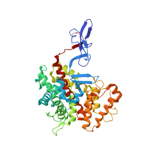
The citrate synthase (CS) of Escherichia coli is an allosteric hexameric enzyme specifically inhibited by NADH. The crystal structure of wild type (WT) E. coli CS, determined by us previously, has no substrates bound, and part of the active site is in a highly mobile region that is shifted from the position needed for catalysis. The CS of Acetobacter aceti has a similar structure, but has been successfully crystallized with bound substrates: both oxaloacetic acid (OAA) and an analog of acetyl coenzyme A (AcCoA). We engineered a variant of E. coli CS wherein five amino acids in the mobile region have been replaced by those in the A. aceti sequence. The purified enzyme shows unusual kinetics with a low affinity for both substrates. Although the crystal structure without ligands is very similar to that of the WT enzyme (except in the mutated region), complexes are formed with both substrates and the allosteric inhibitor NADH. The complex with OAA in the active site identifies a novel OAA-binding residue, Arg306, which has no functional counterpart in other known CS-OAA complexes. This structure may represent an intermediate in a multi-step substrate binding process where Arg306 changes roles from OAA binding to AcCoA binding. The second complex has the substrate analog, S-carboxymethyl-coenzyme A, in the allosteric NADH-binding site and the AcCoA site is not formed. Additional CS variants unable to bind adenylates at the allosteric site show that this second complex is not a factor in positive allosteric activation of AcCoA binding.
Organizational Affiliation:
Department of Chemistry, University of Manitoba, Winnipeg, MB R3T 2N2, Canada.