4J3P

Crystal structure of full-length catechol oxidase from Aspergillus oryzae


Experimental Data Snapshot

  • Method: X-RAY DIFFRACTION
  • Resolution: 2.50 Å
  • R-Value Free: 0.245 
  • R-Value Work: 0.188 
  • R-Value Observed: 0.191 

wwPDB Validation   3D Report Full Report


Ligand Structure Quality Assessment 


This is version 2.1 of the entry. See complete history


Literature

The crystal structure of an extracellular catechol oxidase from the ascomycete fungus Aspergillus oryzae.

Hakulinen, N.Gasparetti, C.Kaljunen, H.Kruus, K.Rouvinen, J.

(2013) J Biol Inorg Chem 18: 917-929

  • DOI: https://doi.org/10.1007/s00775-013-1038-9
  • Primary Citation of Related Structures:  
    4J3P, 4J3Q, 4J3R

  • PubMed Abstract: 

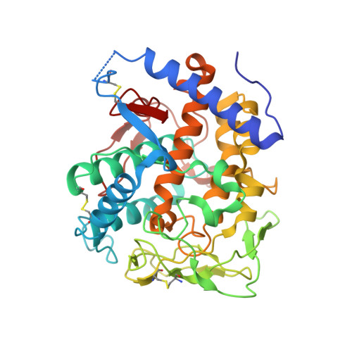
    Catechol oxidases (EC 1.10.3.1) catalyse the oxidation of o-diphenols to their corresponding o-quinones. These oxidases contain two copper ions (CuA and CuB) within the so-called coupled type 3 copper site as found in tyrosinases (EC 1.14.18.1) and haemocyanins. The crystal structures of a limited number of bacterial and fungal tyrosinases and plant catechol oxidases have been solved. In this study, we present the first crystal structure of a fungal catechol oxidase from Aspergillus oryzae (AoCO4) at 2.5-Å resolution. AoCO4 belongs to the newly discovered family of short-tyrosinases, which are distinct from other tyrosinases and catechol oxidases because of their lack of the conserved C-terminal domain and differences in the histidine pattern for CuA. The sequence identity of AoCO4 with other structurally known enzymes is low (less than 30 %), and the crystal structure of AoCO4 diverges from that of enzymes belonging to the conventional tyrosinase family in several ways, particularly around the central α-helical core region. A diatomic oxygen moiety was identified as a bridging molecule between the two copper ions CuA and CuB separated by a distance of 4.2-4.3 Å. The UV/vis absorption spectrum of AoCO4 exhibits a distinct maximum of absorbance at 350 nm, which has been reported to be typical of the oxy form of type 3 copper enzymes.


  • Organizational Affiliation

    Department of Chemistry, University of Eastern Finland, Joensuu Campus, PO Box 111, 80101, Joensuu, Finland, nina.hakulinen@uef.fi.


Macromolecules
Find similar proteins by:  (by identity cutoff)  |  3D Structure
Entity ID: 1
MoleculeChains Sequence LengthOrganismDetailsImage
Catechol oxidase383Aspergillus oryzaeMutation(s): 0 
Gene Names: 5990879
EC: 1.10.3.1
UniProt
Find proteins for Q2UNF9 (Aspergillus oryzae (strain ATCC 42149 / RIB 40))
Explore Q2UNF9 
Go to UniProtKB:  Q2UNF9
Entity Groups  
Sequence Clusters30% Identity50% Identity70% Identity90% Identity95% Identity100% Identity
UniProt GroupQ2UNF9
Sequence Annotations
Expand
  • Reference Sequence
Oligosaccharides

Help

Entity ID: 2
MoleculeChains Length2D Diagram Glycosylation3D Interactions
beta-D-mannopyranose-(1-4)-2-acetamido-2-deoxy-beta-D-glucopyranose-(1-4)-2-acetamido-2-deoxy-beta-D-glucopyranose
B
3N-Glycosylation
Glycosylation Resources
GlyTouCan:  G15407YE
GlyCosmos:  G15407YE
GlyGen:  G15407YE
Small Molecules
Ligands 5 Unique
IDChains Name / Formula / InChI Key2D Diagram3D Interactions
NAG
Query on NAG

Download Ideal Coordinates CCD File 
F [auth A],
G [auth A],
H [auth A]
2-acetamido-2-deoxy-beta-D-glucopyranose
C8 H15 N O6
OVRNDRQMDRJTHS-FMDGEEDCSA-N
MAN
Query on MAN

Download Ideal Coordinates CCD File 
I [auth A]alpha-D-mannopyranose
C6 H12 O6
WQZGKKKJIJFFOK-PQMKYFCFSA-N
SO4
Query on SO4

Download Ideal Coordinates CCD File 
J [auth A]SULFATE ION
O4 S
QAOWNCQODCNURD-UHFFFAOYSA-L
CU
Query on CU

Download Ideal Coordinates CCD File 
C [auth A],
D [auth A]
COPPER (II) ION
Cu
JPVYNHNXODAKFH-UHFFFAOYSA-N
OXY
Query on OXY

Download Ideal Coordinates CCD File 
E [auth A]OXYGEN MOLECULE
O2
MYMOFIZGZYHOMD-UHFFFAOYSA-N
Experimental Data & Validation

Experimental Data

  • Method: X-RAY DIFFRACTION
  • Resolution: 2.50 Å
  • R-Value Free: 0.245 
  • R-Value Work: 0.188 
  • R-Value Observed: 0.191 
  • Space Group: P 32 2 1
Unit Cell:
Length ( Å )Angle ( ˚ )
a = 118.88α = 90
b = 118.88β = 90
c = 84.49γ = 120
Software Package:
Software NamePurpose
MAR345dtbdata collection
SOLVEphasing
PHENIXrefinement
XDSdata reduction
XSCALEdata scaling

Structure Validation

View Full Validation Report


Ligand Structure Quality Assessment 


Entry History 

Deposition Data

Revision History  (Full details and data files)

  • Version 1.0: 2013-11-27
    Type: Initial release
  • Version 1.1: 2013-12-04
    Changes: Database references
  • Version 2.0: 2020-07-29
    Type: Remediation
    Reason: Carbohydrate remediation
    Changes: Atomic model, Data collection, Derived calculations, Structure summary
  • Version 2.1: 2023-09-20
    Changes: Data collection, Database references, Refinement description, Structure summary